P. ramorum photomicrographs

Home

Sporulation Thumbnails   More details

Click on any of these thumbnails to view an enlargement.

Sporangia

Sporangia

Sporangia

Sporulation

Sporangia & chlamydospores

Chlamydospores

Ultrastructural Features Thumbnails
More details

Zoospores

Zoospores

Zoospores

Zoosporangia

Zoospore release

Zoospore release

Zoosporangia

P. ramorum cysts

P. ramorum cyst

Cyst germination

Chlamydospores

Chlamydospores

Chlamydospore

Chlamydospores

Chlamydospore

Hyphae

Lenticel

Hypahe in lenticels

Infected tanoak tissue

Tanoak cortex

Hyphae in xylem

Infected bay leaf

Sporulation Thumbnails: The six sporulation photomicrographs represent a time series (20 hours to 9 days) showing the sporulation of Phytophthora ramorum on bay leaf tissue that was artificially inoculated in the lab.

Photomicrographs by Dr. Jennifer L. Parke
Associate Professor (Research)
Department of Botany and Plant Pathology
& Department of Crop and Soil Science
Oregon State University, Corvallis, OR 97331

To download PDF files for printing, click on the appropriate link:

          Sporangia 1   Sporangia 2   Sporangia 3
          Sporulation   Sporangia & chlamydospores
          Chlamydospores

Back to Top

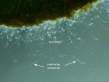
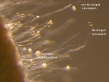
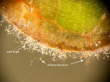
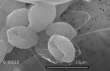
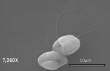
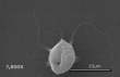
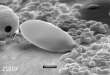
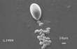
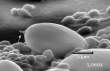
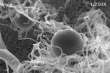
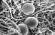
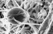
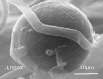
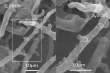
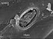
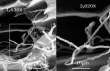
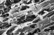
Ultrastructural Features Thumbnails: The 22 ultratructural photomicrographs show the various ultrastructural features of Phytophthora ramorum. If you would like copies of the following images for research or educational purposes, please contact Ed Florance at one of the email addresses listed below.

Edwin R. Florance Ph.D.
Professor Emeritus
Biology Department
Lewis & Clark College
0615 SW Palatine Hill Rd.
Portland, OR 97219
florance@lclark.edu
florance@comcast.net

This work was funded in part by the USDA Forest Service Pacific Southwest Research Station.

Back to Top