
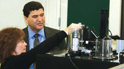
Dr. Carolyn Larabell of Lawrence Berkeley National Laboratory shows NIH Director Dr. Elias Zerhouni one of the features of a new one-of-a-kind microscope that uses X-rays to look inside cells.
Photo courtesy of Lawrence Berkeley National Laboratory
Director Dr. Elias Zerhouni is a world-renowned leader in the field of biomedical imaging—a science that uses advanced technologies to capture, store, analyze, and display images of the body. He is credited with developing imaging methods used for diagnosing cancer and cardiovascular disease. He is also one of the world's premier experts in magnetic resonance imaging (MRI), as well as computed tomographic densitometry.
MedlinePlus: As a leader in biomedical imaging, what do you consider most important for the public to understand about the field today?
Dr. Zerhouni: Biomedical imaging is fulfilling a fundamental dream of scientists to be able to peek into the human body without destroying what we're looking at. That was the impetus for my own research. Before imaging, you had to do surgery. Over the next 40 years, imaging is going to help unravel the mysteries of how our molecules, cells, tissues, and systems interact. It will be important for medical applications, but even more important for basic research and understanding biology.
MedlinePlus: There has been lots of news recently about the value of CAT scanning in detecting certain cancers. What is your view?
Dr. Zerhouni: Essentially, computerized axial tomography (CAT) is a way of looking at biology indirectly; at anatomy, structure, and function. And whenever you have an indirect technique, you want to know how accurate it is. You don't want too many false negatives, where you are missing something, or false positives, where you are seeing something that you think is a disease but isn't.
That's why CAT scanning has been developed—to detect lesions in organs like the lung or the rest of the body. And that's why we're developing other tests, like ultrasound for example, to look at intrauterine pregnancies, or mammography to look at early cancers.
But you have to be very careful. The value of such testing depends on its accuracy. For accuracy, we need to do very rigorous trials. It's not enough to believe that something works. We have to provide the American public with scientific, evidence-based answers.
MedlinePlus: Your work has helped move forward the science of magnetic resonance imaging (MRI). What do you see as its current and long-term benefits?
"My interest, my dream, was that, in fact, you could look inside the human body without destroying it."
Dr. Zerhouni: My work has been directed at the intersection of the physical sciences—mathematics, physics, engineering— and biological sciences. I believe deeply that we must be able to quantify what happens in biology. It's not enough to know that this molecule interacts with that one, or by how much and when? You need to quantify it.
From the beginning, I have sought to bring to biology rigorous quantitative techniques at all levels of imaging, whether trying to diagnose disease in the hospital, or in the laboratory. Quantification is the key to the future. Science advances because it has better tools to measure exactly what happens in the spot where it happens. That's what imaging is all about: to localize or extract information, then do something with it.
MedlinePlus: What sparked your interest in radiology and medical imaging?
Dr. Zerhouni: At the time I became interested, radiology was a backwater—not very prestigious. But I loved physics and math, was interested in medicine, and had an uncle who was a radiologist. He said, "You know, you might be able to marry those two things." One day, he showed me an image of the world's very first CAT scan. It was grainy and terrible. But when he told me how it was acquired—with an X-ray that goes around with a computer recording the data, I said, "This is it. I can contribute to this!" My interest, my dream, was that, in fact, you could look inside the human body without destroying it.
MedlinePlus: You said that we are entering a new era in biomedical imaging. What do you see as its long-term opportunities and benefits?
Dr. Zerhouni: The new era is really the need to understand exactly the complex biological interactions that result in disease—or good health, from the molecular and atomic levels on up. Whether it is electron microscopy or CAT scanning, mouse or human imaging, all are bound by a common thread. And that thread is the quest to know what happens inside, without destroying the system and understanding how it really works.
MedlinePlus: How is NIH prepared to take advantage of this new biomedical-imaging era?
Dr. Zerhouni: We've seen an explosion of new ideas. For example, at NIH, Dr. Jennifer Lippincott-Schwartz and colleagues have created photo-activated light microscopy. This is a technique that relies on digital optical imaging, rather than X-rays, CAT scanning, or MRIs. It can actually highlight a single molecule with another that emits light. And the light can be triggered to determine the exact spot in the cell where and how something happens. This is going to revolutionize the way we understand biology in health and disease.
MedlinePlus: To approach the topic differently, what promising new developments and technologies in imaging can we expect in the near future?
Dr. Zerhouni: Today, as many as 25 different techniques are under way. With magnetic resonance imaging, for example, we can see what regions of the brain are activated when you think or do things. That was an impossible dream 15 years ago that now has enormous impact.
There is also a convergence of chemistry with the development of molecules that, for instance, can shine light or send signals that can be detected with a microscope or an ultrasound machine. So scientists are now thinking, "Wait, if I can see something, then I can do something about it." This is leading to nanotechnology cancer therapy, for example, in which drugs are inserted in a molecule that is homed in on a cancer cell, and then triggered by ultrasound or other techniques, such as heat.
MedlinePlus: Imaging technologies aren't cheap. Will NIH and other imaging pioneers have the funding to fulfill the promise of these technologies in the United States?
Dr. Zerhouni: When I began researching, in 1980, the first MRI scanners cost $3 million—the equivalent of $5 million today. People warned me away, saying "It's going to be a dead end because it's so expensive." Over the years, however, just as with cell phones, the computers have become more powerful, the MRI procedure faster and cheaper.
It's always the same with new technology. The first prototypes are always expensive, but they must be supported. If not, then all that the new field promises will never be explored, let alone fulfilled.
MedlinePlus: Is there anything else?
Dr. Zerhouni: The key in science today is to understand that the complexity of biology is much greater than we thought 20 or 30 years ago, and as we've discovered more, we need more information at the local level. That is why imaging has become so important.
Even though I was fortunate to be in on its birth, I believe we are on the verge of another period in which imaging is going to be even more central to our understanding of biology. I would urge young scientists to get in on this exciting time. But I would also caution that to be good in imaging, they will have to be good at multiple disciplines. And they must be able to work with different people.
Chemistry must work with physics, which has to work with biology, with medicine, surgery, and all that. It may be difficult. But it is exciting. So my message is: break the barriers!