
General Information About Hypopharyngeal Cancer
Key Points for This Section
- Hypopharyngeal cancer is a disease in which malignant (cancer) cells form in the tissues of the hypopharynx.
- Use of tobacco products and heavy drinking can affect the risk of developing hypopharyngeal cancer.
- Possible signs of hypopharyngeal cancer include a sore throat and ear pain.
- Tests that examine the throat and neck are used to help detect (find) and diagnose hypopharyngeal cancer.
- Certain factors affect prognosis (chance of recovery) and treatment options.
Hypopharyngeal cancer is a disease in which malignant (cancer) cells form in the tissues of the hypopharynx.
The hypopharynx is the bottom part of the pharynx (throat). The pharynx is a hollow tube about 5 inches long that starts behind the nose, goes down the neck, and ends at the top of the trachea (windpipe) and esophagus (the tube that goes from the throat to the stomach). Air and food pass through the pharynx on the way to the trachea or the esophagus.
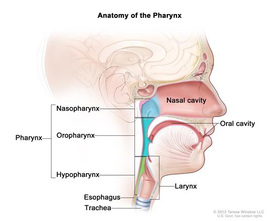
Most hypopharyngeal cancers form in squamous cells, the thin, flat cells lining the inside of the hypopharynx. The hypopharynx has 3 different areas. Cancer may be found in 1 or more of these areas.
Hypopharyngeal cancer is a type of head and neck cancer.
Use of tobacco products and heavy drinking can affect the risk of developing hypopharyngeal cancer.
Risk factors include the following:
- Smoking tobacco.
- Chewing tobacco.
- Heavy alcohol use.
- Eating a diet without enough nutrients.
- Having Plummer-Vinson syndrome.
Possible signs of hypopharyngeal cancer include a sore throat and ear pain.
These and other symptoms may be caused by hypopharyngeal cancer. Other conditions may cause the same symptoms. A doctor should be consulted if any of the following problems occur:
- A sore throat that does not go away.
- Ear pain.
- A lump in the neck.
- Painful or difficult swallowing.
- A change in voice.
Tests that examine the throat and neck are used to help detect (find) and diagnose hypopharyngeal cancer.
The following tests and procedures may be used:
- Physical exam of the throat: An exam in which the doctor feels for swollen lymph nodes in the neck and looks down the throat with a small, long-handled mirror to check for abnormal areas.
- Endoscopy: A procedure used to look at areas in the throat that cannot be seen with a mirror during the physical exam of the throat. An endoscope (a thin, lighted tube) is inserted through the nose or mouth to check the throat for anything that seems unusual. Tissue samples may be taken for biopsy.
- CT scan (CAT scan): A procedure that makes a series of detailed pictures of areas inside the body, taken from different angles. The pictures are made by a computer linked to an x-ray machine. A dye may be injected into a vein or swallowed to help the organs or tissues show up more clearly. This procedure is also called computed tomography, computerized tomography, or computerized axial tomography.
- MRI (magnetic resonance imaging): A procedure that uses a magnet, radio waves, and a computer to make a series of detailed pictures of areas inside the body. This procedure is also called nuclear magnetic resonance imaging (NMRI).
- Head, neck, and chest x-rays: An x-ray of the head, neck, and organs and bones inside the chest. An x-ray is a type of energy beam that can go through the body and onto film, making a picture of areas inside the body.
- Barium esophagogram: An x-ray of the esophagus. The patient drinks a liquid that contains barium (a silver-white metallic compound). The liquid coats the esophagus and x-rays are taken.
- Esophagoscopy: A procedure to look inside the esophagus to check for abnormal areas. An esophagoscope (a thin, lighted tube) is inserted through the mouth or nose and down the throat into the esophagus. Tissue samples may be taken for biopsy.
- Bronchoscopy: A procedure to look inside the trachea and large airways in the lung for abnormal areas. A bronchoscope (a thin, lighted tube) is inserted through the nose or mouth into the trachea and lungs. Tissue samples may be taken for biopsy.
- Biopsy: The removal of cells or tissues so they can be viewed under a microscope to check for signs of cancer.
Certain factors affect prognosis (chance of recovery) and treatment options.
Prognosis (chance of recovery) depends on the following:
- The stage of the cancer (whether it affects part of the hypopharynx, involves the whole hypopharynx, or has spread to other places in the body). Hypopharyngeal cancer is usually detected in later stages because early symptoms rarely occur.
- The patient's age, gender, and general health.
- The location of the cancer.
- Whether the patient smokes during radiation therapy.
Treatment options depend on the following:
- The stage of the cancer.
- Keeping the patient's ability to talk, eat, and breathe as normal as possible.
- The patient's general health.
Patients who have had hypopharyngeal cancer are at an increased risk of developing a second cancer in the head or neck. Frequent and careful follow-up is important.
 Back to Top