
Rabies and Kids!
Electron microscopy
The ultrastructure of viruses can be examined by electron microscopy. Using this method, the structural components of viruses and their inclusions can be observed in detail. Rabies virus is in the family of Rhabdoviruses. When viewed with an electron microscope Rhabdoviruses are seen as bullet-shaped particles.
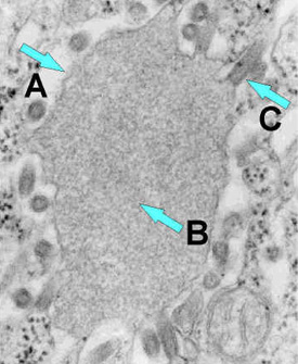
Rabies virus budding from an inclusion (Negri body) into the endoplasmic reticulum in a nerve cell. A = Negri body. B = abundant RNP in the inclusion. C = Budding rabies virus.
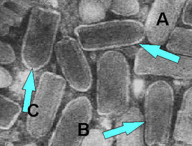
Negatively stained Rhabdovirus as seen through an electron microscope. Notice the bullet shape of the virus (A). See the "bee hive" like striations of the RNP (B). Notice the glycoprotein spikes in the outer member bilayer (C).

Ribonucleoprotein - notice the abundant strands of coiled RNP (almost everything in the image is RNP).
Get email updates
To receive email updates about this page, enter your email address:
Contact Us:
- Centers for Disease Control and Prevention
1600 Clifton Rd
Atlanta, GA 30333 - 800-CDC-INFO
(800-232-4636)
TTY: (888) 232-6348 - New Hours of Operation
8am-8pm ET/Monday-Friday
Closed Holidays - cdcinfo@cdc.gov