National Cancer Institute NCI Cancer Bulletin: A Trusted Source for Cancer Research News
September 18, 2012 • Volume 9 / Number 18

NEWS

Lung Cancer Genome Surveys Find Many Potential Drug Targets

Illustration of a man's upper body showing the lungs

Five new studies have identified genetic and epigenetic alterations in hundreds of lung tumors, including many changes that could be targeted by drugs that are already available or in clinical testing.

The reports, all published this month, included genomic information on more than 400 lung tumors. In addition to confirming genetic alterations previously tied to lung cancer, the studies identified other changes that may play a role in the disease. Read more > >

COMMENTARY

Assistant Secretary for Health Dr. Howard KohChanging Social Norms about Tobacco Use, One Campus at a Time

by Dr. Howard Koh

The assistant secretary for health describes efforts to by the Department of Health and Human Services to reduce tobacco use among young adults.

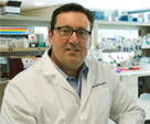
Dr. John Stamatoyannopoulos

A Conversation with Dr. John Stamatoyannopoulos about ENCODE and Cancer Research

The University of Washington oncologist and ENCODE investigator discusses the Encyclopedia of DNA Elements (ENCODE) results and what they could mean for cancer research.


IN DEPTH

UPDATES

  • AHRQ Update

    • Task Force Reaffirms Recommendation against Ovarian Cancer Screening
  • FDA Update

    • FDA Approves Imaging Agent that Helps Detect Prostate Cancer
    • Drug for Advanced Prostate Cancer Approved
    • FDA Approves New Drug to Treat Chronic Myelogenous Leukemia
  • Notes

    • Cancer Classroom Webinar: Culture and Literacy
    • Call for Applications: NCI Clinical Assay Development Program
    • New Brochure Educates Patients about Biospecimen Donation

    MULTIMEDIA

    Containers used in the GTEx PAXgene tissue preservation system (Image from the National Disease Research Interchange GTEx Team)

    Cancer Research Now: Building a Biobank

    Researchers with the NCI cancer Human Biobank describe how donated tissue is collected, processed, shipped, and analyzed for the Genotype-Tissue Expression project.
    Go to Video


Selected articles from past issues of the NCI Cancer Bulletin are available in Spanish.

The NCI Cancer Bulletin is produced by the National Cancer Institute (NCI), which was established in 1937. Through basic, clinical, and population-based biomedical research and training, NCI conducts and supports research that will lead to a future in which we can identify the environmental and genetic causes of cancer, prevent cancer before it starts, identify cancers that do develop at the earliest stage, eliminate cancers through innovative treatment interventions, and biologically control those cancers that we cannot eliminate so they become manageable, chronic diseases.

For more information about cancer, call 1-800-4-CANCER or visit http://www.cancer.gov.

NCI Cancer Bulletin staff can be reached at ncicancerbulletin@mail.nih.gov.

Featured Article

Lung Cancer Genome Surveys Find Many Potential Drug Targets

Illustration of a man’s upper body showing the lungs
New information about genetic changes in lung tumors could help doctors match patients with existing targeted drugs.

Five new studies have identified genetic and epigenetic alterations in hundreds of lung tumors, including many changes that could be targeted by drugs that are already available or in clinical testing.

The reports, all published this month, included genomic information on more than 400 lung tumors. In addition to confirming genetic alterations previously tied to lung cancer, the studies identified other changes that may play a role in the disease. (Links to the study abstracts appear in the sidebar below.)

“These five papers are the first major salvo of genome-wide studies using all of the newest technologies to analyze a large number of lung cancers,” said Dr. John Minna, a clinician and lung cancer researcher at the University of Texas Southwestern Medical Center, who co-authored one of the studies.

Collectively, the studies covered the main forms of the disease—lung adenocarcinomas, squamous cell cancers of the lung, and small cell lung cancers.

Although preliminary, the findings could be used to develop molecular markers for identifying patients who are candidates for certain targeted drugs. At the same time, researchers in the lab can now evaluate the newly discovered changes to identify novel potential therapeutic targets.

“All of these studies say that lung cancers are genomically complex and genomically diverse,” said Dr. Matthew Meyerson of Harvard Medical School and the Dana-Farber Cancer Institute, who co-led several of the studies, including a large-scale analysis of squamous cell lung cancer by The Cancer Genome Atlas (TCGA) Research Network.

Some genes, Dr. Meyerson noted, were inactivated through different mechanisms in different tumors. He cautioned that little is known about alterations in DNA sequences that do not encode genes, which is most of the human genome.

Squamous Cell Tumors

The TCGA investigators sequenced the genomes or exomes (the protein-coding regions of DNA) of tumor samples from 178 patients with squamous cell lung cancer. In more than half of the tumors examined, the researchers found a change to a gene or a signaling pathway that is targeted by a drug that exists or is in development. The findings were reported in Nature on September 9.

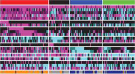
Figure showing squamous lung cancer tumors grouped by gene expression subtype
Detail of a figure showing gene-expression differences in subtypes of squamous cell lung cancer

“This gives us an enormous opportunity for progress in this disease,” said Dr. Meyerson.

The TCGA model integrates genomic data for squamous cell lung cancers with clinical information, when available, and with other tumor characteristics, such as gene expression, epigenetic changes to cells, and alterations in the number of gene copies.

“The framework for these five studies was built on a convergence of new technologies and the need to better understand the biology of lung cancers as it relates to new therapies for our patients,” said Dr. Paul Paik, who treats patients with lung cancer at Memorial Sloan-Kettering Cancer Center and was part of the clinical team involved in TCGA.

Small studies (for example, here and here) have provided hints that certain signaling pathways are important in squamous cell lung cancers, leading to new trials of targeted drugs. “Now, with the publication of the TCGA study, we know that squamous cell lung cancers have a myriad of potentially targetable changes,” Dr. Paik noted.

An unexpected finding was the presence of mutations in the EGFR gene in about 1 percent of squamous cell tumors. These tumors might respond to available drugs that block signals through the EGFR pathway.

The researchers also found evidence of genetic changes that may help lung cancer cells evade surveillance by the immune system.

Testing Lung Tumors

Any therapeutic targets to emerge from the new reports would need to be incorporated into molecular tests that can identify candidates for certain drugs. A leader in this work is the Lung Cancer Mutation Consortium, which has been building knowledge of the mutations associated with the disease and testing for these changes.

Many patients with lung adenocarcinomas have benefited from targeted drugs. Crizotinib (Xalkori), for instance, has elicited some dramatic responses in patients whose tumors harbor a particular gene fusion. Some medical centers now routinely test tumors before selecting treatment for patients with lung adenocarcinomas.

“If you look at lung cancer as a whole, the big therapeutic targets were first identified in adenocarcinomas,” Dr. Minna explained. “Now there are new targeted therapies that could make an impact on squamous cell lung cancer.”

At Memorial Sloan-Kettering, all patients with squamous cell lung cancer have their tumors tested for drug targets using various techniques, including DNA sequencing. Among 28 of these patients evaluated recently, more than 60 percent had tumors that contained a potential target.   

Dr. Paik noted that his group will use the TCGA results to expand their testing. “In a sense, the future potential of this new work is being realized now,” he said. “That’s pretty exciting.”

Small Cell Lung Cancer

Two new reports describe genetic changes in small cell lung cancers, which tend to be aggressive and about which little has been known. The research teams conducted exome or whole-genome sequencing on a total of 82 samples of such tumors.

“This study gave us a host of new targets to explore,” said Dr. Charles Rudin of the Johns Hopkins Kimmel Cancer Center, who led one study. The next steps will be to validate which targets are driving the growth of tumors and are “druggable,” he added.

The researchers found that a gene called SOX2, which plays a role in normal development, may contribute to some small cell lung cancers, as well as other cancers, and could be targeted.

Small cell lung cancers have been challenging to study because most are not treated surgically, so tumor samples are rare. What’s more, these tumors have high rates of genetic mutations due to tobacco smoke, yet only some mutations are driving the disease, noted Dr. Roman Thomas of the University of Cologne in Germany, who led the other study.

Using statistical “filters,” his group found that genes involved in modifying histone proteins, which help package DNA within a cell, were frequently mutated in the disease.

“These cancers are extraordinarily complex, so as researchers our steps forward are incremental—but, still, they are steps,” Dr. Thomas noted. “No one would have imagined that lung cancer would be the prototypical disease for targeted medicine.”

Comparing Tumors in Smokers and Nonsmokers

Non-small cell lung cancers were the focus of two additional studies, which appeared in Cell. One group sequenced the exomes or genomes of 183 tumor samples, and the other conducted whole-genome sequencing of tumor tissues from 17 smokers and nonsmokers.

“We found a substantially lower number of mutations in the genomes of tumors from nonsmokers compared to the smokers,” said Dr. Ramaswamy Govindan of the Washington University School of Medicine in St. Louis, MO, who led the study. Five study participants who had never smoked had a mutation that could be targeted by an existing drug.

All these studies show how diverse and how complicated the cancer genome is. But we now have a panoramic view of the genomic landscape, and this is important for moving forward in this disease.

—Dr. Ramaswamy Govindan
In all, the study authors found 54 genes with potentially targetable alterations in the 17 patients.

“The days of large clinical trials for lung cancer are over,” Dr. Govindan said, noting that patients need to be selected for specific treatments based on the characteristics of their tumors. “We also need to develop clinical trials that move targeted therapies to earlier stages of lung cancer, where we have a better chance of a cure.”

Future clinical trials, he predicted, would look for relatively large effects of drugs in selected patients. Dr. Minna agreed, saying, “If the effects are not there, we will move on to the next target and the next drug.”

The new results are really a teaser for what’s coming. TCGA plans to sequence a total of 500 adenocarcinomas and 500 squamous cells tumors. These results could help shed light on issues such as epigenetic changes in lung cancer, mechanisms of drug resistance, and how tumors are influenced by the surrounding tumor microenvironment.

“All these studies published back to back show how diverse and how complicated the cancer genome is,” Dr. Govindan said. “But we now have a panoramic view of the genomic landscape, and this is important for moving forward in this disease.”

Dr. Minna added, “After treating thousands of patients with lung cancer and not doing too well, I am very excited about the new results.”

Edward R. Winstead

The articles mentioned in this story were funded in part by NIH (01GS08100, 01GS08101, CA140594, P30 CA021765, P50 CA058187, P50 CA090578, P50 CA058184, P50 CA119997, P50 CA70907, R01 CA122794 , T32 CA9216, T32 GM07753, U24 CA126543, U24 CA126544, U24 CA126546, U24 CA126551, U24 CA126554, U24 CA126561, U24 CA126563, U24 CA143799, U24 CA143835, U24 CA143840, U24 CA143843, U24 CA143845, U24 CA143848, U24 CA143858, U24 CA143866, U24 CA143867, U24 CA143882, U24 CA143883, U24 CA144025, U54 HG003067, U54 HG003079, U54 HG003079, U54 HG003273).

Cancer Research Highlights

Diagnostic Radiation Exposure May Raise Breast Cancer Risk in Some BRCA1/2 Mutation Carriers

Radiation from conventional x-rays, mammograms, and other diagnostic tests before age 30 may increase the risk of breast cancer in women who carry BRCA1 or BRCA2 gene mutations. In a large, retrospective cohort study, this increased risk was seen at radiation doses considerably lower than those associated with increased risk of breast cancer in other cohorts exposed to radiation.

The findings, from an analysis of women in France, the Netherlands, and the United Kingdom who participated in the GENE-RAD-RISK study, were reported September 6 in the British Medical Journal.

Exposure to ionizing radiation is an established risk factor for breast cancer, particularly when the exposure occurs at an early age. Because the BRCA1 and BRCA2 proteins are important in repairing DNA damage, including damage caused by radiation, researchers have hypothesized that carriers of a mutation in one of these genes might be more sensitive than the general population to ionizing radiation. But previous studies designed to answer this question have yielded inconsistent results.

The new study, led by Dr. Flora van Leeuwen of the Netherlands Cancer Institute in Amsterdam, focused on 1,122 women aged 18 or older who were known to carry a BRCA1 or BRCA2 mutation. The women reported their histories of all diagnostic procedures involving radiation to the chest or shoulders. Researchers used this information to estimate the cumulative dose of radiation to the breast for each woman. They then used national registries or medical records to confirm breast cancer diagnoses among the participants.

When compared with no exposure, any exposure to diagnostic radiation before age 30 was associated with almost double the risk of breast cancer in BRCA1/2 mutation carriers. Women who received the highest doses of radiation before age 30 had an almost fourfold higher risk. By contrast, there was no evidence of an increased breast cancer risk associated with exposure at ages 30 to 39.

Because women with BRCA1 or BRCA2 mutations have a greatly increased risk of developing breast cancer, some guidelines recommend annual mammograms beginning at age 25 to 35 for these women.

These findings “support the recommendation to use non-ionising radiation imaging techniques (such as MRI) as the main tool for surveillance in young BRCA1 and BRCA2 mutation carriers,” the authors concluded.

Dr. Barry Kramer, director of NCI’s Division of Cancer Prevention, noted that the study “helps refine our knowledge” about the connection between radiation exposure and breast cancer risk in people who may be particularly sensitive to the effects of ionizing radiation. However, he cautioned, “the evidence in this study is not definitive.”

The authors acknowledge that their reliance on self report of prior radiation exposure could introduce statistical bias. “People who have cancer may be more likely than those without cancer to recall any radiation that they had,” Dr. Kramer explained.

Until more definitive evidence is available, he continued, “Women who carry BRCA1 or BRCA2 mutations ought to know what we do and don’t know.” Physicians should review the screening and prevention options with each woman and help her consider the potential benefits and downsides of each, he concluded.

Further reading: “Clinical Management of BRCA Mutation Carriers

New Drug Improves Survival in Patients with Advanced Lung Cancer

UPDATE

The September 18, 2012, issue of the NCI Cancer Bulletin included a Cancer Research Highlight reporting the results of a phase II trial that tested the experimental drug bavituximab in patients with non-small cell lung cancer. The results were presented at the 2012 Chicago Multidisciplinary Symposium in Thoracic Oncology.

On September 24, Peregrine Pharmaceuticals, the company that manufactures bavituximab and sponsored the phase II clinical trial, issued a news release explaining that the company had identified “major discrepancies” with some aspects of how the trial was conducted. The company intends to communicate further as soon as it determines the impact of the discrepancies.

Drug Targeting Tumor Suppressor Shows Promise in First Human Study

An experimental drug that reactivates mutant forms of the tumor suppressor protein p53 is safe for humans, according to results from a phase I trial. The drug, APR-246, also stimulated signaling pathways that control p53 in tumor cells isolated from peripheral blood.

The study, published in the Journal of Clinical Oncology, was led by Dr. Sören Lehmann of Karolinska University Hospital in Stockholm.

The findings represent “a major step forward in targeting the most frequently altered pathway in cancer,” wrote Drs. Brian D. Lehmann and Jennifer A. Pietenpol of Vanderbilt University School of Medicine in an accompanying article.

The p53 protein suppresses tumor growth by increasing the expression of genes that slow the cell cycle, that prevent cells from dividing, or that cause programmed cell death (apoptosis). At least half of all tumors develop inactivating mutations in TP53, the gene that produces p53, allowing the tumor to evade this regulation. APR-246 counteracts TP53 mutations by restoring the gene-regulatory activity of mutant p53 protein and by inducing the death of cancer cells.

The 22 participants enrolled in the trial had various forms of leukemia, hormone-refractory metastatic prostate cancer, non-Hodgkin lymphoma, or multiple myeloma. These patients were included because prostate cancers have a high rate of TP53 mutations and because, in preclinical studies, leukemia cells were particularly sensitive to drugs that target p53.

The patients received intravenous infusions of APR-246 for 4 consecutive days. After a follow-up period of 17 days, the most common side effects were fatigue, dizziness, headache, and confusion.

While the trial was not designed to assess the drug’s antitumor effects, several patients showed clinical responses. To investigate the biologic activity of APR-246, Dr. Lehmann and his colleagues analyzed circulating tumor cells obtained before and after the 4-day treatment from the six patients who had these cells. Four of these patients had fewer proliferating cells after receiving APR-246. The investigators also observed signs of apoptosis and increased expression of several p53 target genes.

Microarray analysis of RNA isolated from the circulating tumor cells showed changes in genes responsible for regulating cell growth and death. The level of expression of genes that promote apoptosis appeared to correlate with the dose of APR-246.

Trials are being designed to investigate the effects of higher exposures to APR-246 through longer infusion times and of combining APR-246 with other chemotherapy drugs, most of which depend on a functional version of p53 to be effective.

Study Reveals How Breast Cancer Spreads to Lymph Nodes

A recent study offers new insights into how breast cancer may spread to nearby lymph nodes and also suggests that a drug commonly used to treat heart failure, digoxin, may be able to interrupt the process. The findings were reported September 10 in the Proceedings of the National Academies of Sciences (PNAS).

The lymphatic system is an important route through which cancer cells reach the circulatory system and travel to distant organs, where they develop into metastatic tumors. Metastasis—which is responsible for most cancer deaths—is not well understood, and few treatments actively target it. In breast cancer, nearly all women with metastatic disease have lymph node involvement.

Using mice, Dr. Gregg Semenza of Johns Hopkins University and his colleagues showed that hypoxia-inducible factor-1 alpha (HIF-1α) plays a direct role in the spread of breast cancer cells to the lymph nodes. HIF-1 α is a subunit of the HIF-1 protein, which promotes blood vessel formation under low-oxygen conditions, such as those in tumors. HIF-1α, the researchers found, activates the PDGF-B gene, which codes for platelet-derived growth factor (PDGF-B).

When mice with tumors formed from injected human breast cancer cells were treated with digoxin (which inhibits HIF-1α) or with imatinib (Gleevec; which inhibits PDGF-B), cancer cell spread was dramatically reduced.

In addition, mice with tumors formed from breast cancer cells that were genetically modified to block production of HIF-1α had 75 percent fewer lymph node metastases than mice with tumors formed from unmodified breast cancer cells.

In biopsy samples from human breast cancers, the authors also found that 

  • PDGF-B was highly active in cells that were starved of oxygen, 
  • HIF-1α directly activated transcription of PDGF-B,
  • the HIF-1α and PDGF-B proteins were found near each other in almost all of the biopsy samples they studied, and 
  • levels of expression of these proteins in biopsy samples correlated with tumor grade.

Other studies have linked HIF-1α and PDGF-B to metastatic spread. “But this is the first time that anybody has connected all the dots in a single cancer,” Dr. Semenza explained.

Later this year, the Hopkins researchers plan to launch an early-phase clinical trial to test digoxin in women with operable breast cancer, Dr. Semenza said. Digoxin, an off-patent drug, will be given for about 2 weeks before surgery, and the researchers will analyze pre- and post-surgical tumor samples to determine whether the drug is inhibiting HIF-1 and its downstream target genes.

If the trial suggests that the drug is having the intended molecular effects, an early-phase trial combining digoxin with other standard treatments would likely be launched.

Also in the Journals: Smoky Coal Associated with Increased Risk of Lung Cancer in Rural China

Results from a large retrospective cohort study conducted in China suggest that use of smoky (bituminous) coal substantially increases the lifetime risk of developing lung cancer. The findings appeared August 29 in the British Medical Journal.

Researchers compared deaths from lung cancer between lifelong users of smoky coal and of smokeless (anthracitic) coal for cooking and heating in Xuanwei County, Yunnan Province. More than 37,000 people were followed from 1976 to 1996, and more than 2,000 deaths from lung cancer were recorded during that time.

After accounting for other risk factors, including tobacco use, users of smoky coal had a 30-fold greater risk of developing lung cancer than users of smokeless coal. The absolute risk of lung cancer death before age 70 among smoky coal users was 18 percent for men and 20 percent for women, with most of these deaths in nonsmokers. By contrast, the risk was less than 0.5 percent among users of smokeless coal.
 
“The risks from smoky coal are almost as high as those reported for heavy smoking in Western countries and likely represent one of the strongest effects of environmental pollution reported for cancer risk,” said co-author Dr. Qing Lan of NCI’s Division of Cancer Epidemiology and Genetics.

This study was supported in part by NCI's Intramural Research Program.

Also in the Journals: Best Practices Can Reduce Central-Line Infections in Children with Cancer

By using best practices, health providers at Johns Hopkins Children’s Center reduced central line-associated bloodstream infections by 20 percent over 2 years, according to a study published in Pediatrics.

Hospitalized cancer patients often have a central venous access catheter, or central line, inserted into a major blood vessel as a portal for medication, fluids, or blood draws. These central lines can also introduce infection; children with cancer whose treatment has suppressed their immune systems are particularly vulnerable.

The best practices included improved disinfection and a daily assessment to determine whether each central line was needed, could be consolidated, and/or eliminated. In addition, care teams met monthly to discuss the root cause of each central line infection. Family members also received wallet cards describing the correct care of central lines and were encouraged to observe their child’s care and to report any inconsistencies with the best practices.

This study was funded in part by the National Institutes of Health (5Kl2RR025006).

Guest Commentary by Dr. Howard K. Koh

Changing Social Norms about Tobacco Use, One Campus at a Time

Assistant Secretary for Health Dr. Howard Koh

As the Assistant Secretary for Health, I have the honor of advancing a broad portfolio of public health issues on behalf of the Department of Health and Human Services (HHS). An overriding priority is reinvigorating our national commitment to tobacco control. The first-ever HHS Strategic Action Plan for Tobacco Control, entitled Ending the Tobacco Epidemic: A Tobacco Control Strategic Action Plan, commits the department to mobilizing leadership to encourage proven, pragmatic, and achievable interventions at the federal, state, and community levels.

Among other things, the action plan commits to reducing the initiation of tobacco use among young adults, a topic with special relevance to institutions of higher learning. Furthermore, the 31st Surgeon General’s Report on Tobacco, released in March, highlighted some startling statistics pertinent to this goal. Preventing Tobacco Use among Youth and Young Adults notes that 90 percent of all smokers start before age 18, and 99 percent start before age 26. Of concern, progression from occasional to daily smoking frequently occurs during the initial years following high school. Indeed, the number of smokers who initiated smoking after age 18 has increased substantially over the past decade—from 600,000 in 2002 to 1 million in 2010.

The report cites reasons for these disturbing trends. Tobacco industry expenditures related to marketing, promotion, and advertising of tobacco products exceed $1 million per hour—totaling more than $27 million a day. Targeted messages and images portray tobacco use as a desirable and appealing activity. As a result, smoking represents the current social norm in many movies, video games, websites, and communities, thereby promoting a culture that fosters tobacco dependence and disease.

Restoring the social norm to one that, instead, promotes wellness and health requires a commitment to smoke-free and tobacco-free environments.

The Affordable Care Act, the health care law of 2010, is also part of our comprehensive approach toward turning this goal into reality. Most health plans must now cover—without cost-sharing—tobacco-use screening and interventions for tobacco users. The law also makes it easier and more affordable for young adults to get health insurance coverage, by allowing them to stay on their parents’ employer-sponsored or individually purchased health plans.

SmokefreeTXT adSmokefree Teen, a website specifically developed to help teen smokers quit, offers several social media pages to connect teens with cessation tools.

In particular, colleges and universities can take the next step in protecting the health of their students and inspiring change through the adoption of smoke-free and tobacco-free campuses.

To launch a new chapter in ending the epidemic of smoking, I was honored to participate last week in the announcement of the Tobacco-Free College Campus Initiative (TFCCI). The University of Michigan School of Public Health in Ann Arbor hosted the September 12 event, which was webcast to nearly 500 attendees across the country. The TFCCI represents a public/private partnership involving key leaders from universities, colleges, and the public health community to promote the adoption of tobacco-free policies at institutions of higher learning. This landmark public health initiative will protect students, staff, and faculty against involuntary exposure to secondhand smoke while encouraging a change in social norms that can help reduce tobacco use.

To date, more than 700 colleges and universities, representing an estimated 17 percent of institutions of higher learning nationwide, have committed to smoke-free or tobacco-free campus policies.

HHS is pleased to recognize the leadership of institutions that promote public health in this way. Such actions exemplify a key pillar of the tobacco control action plan—“leading by example.” In fact, by adopting a tobacco-free campus policy on July 1, 2011, HHS has already joined the ranks of such institutions leading by example. This action now protects the health of our 80,000 employees who work in dozens of buildings, grounds, and facilities across the country. 

It is my hope that the launch of the TFCCI will encourage all institutions of higher learning to take action. It is time for us to end the epidemic leading to the single most preventable cause of death in this nation. 

Together we can make smoking history.

Dr. Howard K. Koh
Assistant Secretary for Health
U.S. Department of Health and Human Services

A Conversation With

A Conversation with Dr. John Stamatoyannopoulos about ENCODE and Cancer Research

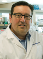
Dr. John Stamatoyannopoulos

For nearly a decade, researchers with the ENCODE (Encyclopedia of DNA Elements) project have been identifying "functional elements” in the human genome sequence—that is, the bits of DNA that help the genome do its job. Recently, the researchers summarized their work in more than 30 published articles. Dr. John Stamatoyannopoulos, an oncologist at the University of Washington in Seattle and ENCODE investigator since the project's inception, spoke with the NCI Cancer Bulletin about what the results could mean for cancer research.

What are the goals of ENCODE?

When ENCODE began in 2003, the overall goal was to annotate all the functional components of the human genome, with an emphasis on the regions that regulate genes. Genes occupy about 2 percent of the genome. And by genes I mean the regions of the genome that get transcribed into RNA and then turned into the proteins that make all the tissues of the body, such as skin and muscle and hair.

Hidden in the remaining 98 percent of the genome are the instructions that tell the genes how to switch on and off in different kinds of cells. A chief goal of ENCODE has been to find those instructions and understand how they are written in the genome.

What do the instructions look like?

In essence, these instructions are organized into millions of DNA “switches.” These switches consist of strings of genetic letters, maybe 100 to 200 letters long, that can be thought of as sentences made up of short DNA words. The DNA words function as docking sites for special regulatory proteins.

When the proteins in a switch are docked to their respective words, the switch becomes active. We have identified, so far, around 4 million switches in the human genome, and there are undoubtedly many more that have not been seen yet.

What was known about regulatory DNA when the project began?

At that time it was well known that genes have switches that control their activity. Up until 2003, perhaps a few hundred switches had been identified. But nobody knew how much of the genome sequence was actually used for things like these switches. Nobody knew how many switches were out there or how many it took to control a gene.

How much of the human genome was thought to be useful?

Cancer cells are taking advantage of the genome’s control circuitry in subtle but important ways that were not previously anticipated.

—Dr. John Stamatoyannopoulos

At the time the project began, the widely quoted estimate was that about 5 percent of the human genome was actually good for something. The outcome of the ENCODE project so far shows that this number was a significant underestimate. As summarized in a paper by ENCODE investigators in Nature, approximately 80 percent of the genome yields a specific biochemical activity, such as the production of RNA or interactions between proteins and DNA.

So, it appears that an extraordinary amount of the genome is used for something. A significant component of this is the switches for controlling genes. Altogether, at least 40 percent of the genome appears to encode such switches, which is a lot!

By comparison, around 5 percent of the genome encodes the information that specifies proteins or the core building blocks of so-called non-coding or RNA genes. And there are many other classes of functional elements, some of which we haven’t yet studied.

What have you learned that is relevant to cancer?

First of all, cancer cells have specialized regulatory DNA switches that we, thus far, do not see in normal cells. We don’t know how to read the information in these switches completely yet, but it should help us decipher the regulatory pathways that are active in cancer cells but not normal cells. And in the past, when we have been able to understand cellular pathways in detail, new types of treatments have followed. This is a very exciting avenue that will be aggressively pursued by many people.

A second interesting aspect of this work relates to studies of genetic variation that has been linked to various cancers through genome-wide association studies. To date, researchers have identified a few hundred genetic variants—single letter DNA changes—associated with the risk for any of 17 kinds of cancer. But 95 percent of these genetic variants do not map to genes. In fact, we reported in Science that the majority of these variants lie in the switches that control genes.

What struck you about the work published so far?

Cancer cells use the genome in ways that we did not anticipate. The results to date show that cancer cells take advantage of the genome’s control circuitry in subtle but important ways that were not previously anticipated. A further understanding of this will deepen our knowledge of what makes a cancer cell tick.

Cancer cells also seem to use special sets of instructions already written into the genome that likely were there originally for some other purpose. Nonetheless, these cancer cells have somehow figured out how to use the instructions. We can now try to understand how this happens.

Has the project changed your thinking about genes?

Yes. It’s clear that we need to think now in terms of groups or networks of genes—of maybe dozens or hundreds of genes, all functioning together. This whole idea of looking at individual genes that are responsible for this or for that is becoming outdated.

How will the results help cancer researchers?

The immediate effect could be to greatly accelerate basic research projects that are already under way. For example, many researchers want to know how a particular gene is regulated, but this typically requires costly and time-consuming experiments. Now, for a huge number of genes researchers will be able to just look this information up online using a viewing application called a genome browser. This is particularly true for genes in cancer cells, because ENCODE used many cancer cell lines.

Ultimately, I think the results will enable a more sophisticated understanding of how cancer cells function relative to normal cells, and this should improve our ability to devise strategies to fight the disease.

This will take time?

Yes. It is important for people to understand the timeline.

We hope these results will accelerate the overall research timetable by helping researchers discover new things more quickly, but nobody should expect that this is going to change clinical practice in the next couple of years.

What is the next phase of your ENCODE research?

We’ve learned that many of these functional elements are highly specialized for different kinds of cells. So creating a complete catalog will require the study of a great many different kinds of cell types and cell states—only a fraction of which have been covered to date. Most of these are going to be normal cells.

And you will continue to study cancer cells as well?

Yes. ENCODE has studied nearly 30 different cancer cell lines so far, but there are obviously many different kinds of cancers; it’s an incredibly interesting area because cancer cells seem to be unique relative to one another as well as relative to normal cells. One of the key advantages of a project like ENCODE is that we gain valuable insights about abnormal cells by making comparisons with a deep catalog of genomic elements that function in normal cells.

The more we study the genome the more interesting things we find. I don’t think anybody should write off how much information is really there.

Interviewed by Edward R. Winstead

Spotlight

Building a Biobank to Explore Mysteries of the Genome

A technician dissects and preserves a tissue sample using the GTEx PAXgene tissue preservation system. (Image from the National Disease Research Interchange GTEx Team)
GTEx is collecting multiple tissue samples from an estimated 1,000 individual donors for genetic research. (Image from the National Disease Research Interchange GTEx Team)

The architects of the biobank wanted nothing left to chance and everything well documented.

That’s why they developed 150 standard operating procedures to ensure that tissue samples were collected, processed, and stored in exactly the same way. And that’s why they are collecting data on the best temperatures for shipping the samples across the United States.

All that planning is paying off for the Genotype-Tissue Expression (GTEx) project, which will use the samples to investigate how genes are regulated in health and disease. Sponsored by the National Institutes of Health (NIH), the project has nearly 4,400 samples of “normal” human tissue from about 175 donors. By collecting many more samples, the project aims to be a resource for studying genetic variation and the regulation of genes in specific tissues.

“This project is an attempt to understand how normal genetic variation influences the expression of genes throughout the body,” said the study’s leader, Dr. Jeffery Struewing of the National Human Genome Research Institute. NCI and the National Institute of Mental Health are also playing lead roles in the effort.

Normal tissue—that is, tissue with no signs of disease—is not routinely collected for research. GTEx is the first large-scale project to collect high-quality samples of up to 32 tissue types from many individual donors.

During a pilot study that began last year, investigators with NCI’s cancer Human Biobank (caHUB) and their collaborators were responsible for acquiring and managing the biospecimens. On average, each organ and tissue donor has contributed 25 types of postmortem tissue, including heart, muscle, and skin. Surgical donors have also contributed tissues.

You will need Adobe Flash Player 8 or later and JavaScript enabled to view this video.

Video produced and edited by Rachael Strecher

A Bigger Biobank

Based on the success of the pilot, the NIH Common Fund is scaling up GTEx, with a goal of reaching 1,000 donors in 3 years. This larger number of donors and samples will provide the statistical power that is needed to ask fundamental questions about the genome, the researchers said.

All cells in the human body contain essentially the same complement of genes, but these genes are activated, or expressed, differently in different types of cells. For the first time, GTEx will allow researchers to investigate how common genetic variants influence the regulation of gene expression using a set of reference tissues.

The relationship between genetic variants and gene regulation in different tissues is “a fascinating biology question, but it is also relevant in medicine,” said Dr. Barbara Stranger of Harvard Medical School and Brigham and Women’s Hospital, who studies genes and complex diseases but is not involved in GTEx.

“Healthy individuals will have some of the same regulatory processes as people with a complex disease,” Dr. Stranger continued. Understanding these processes could provide information about a variety of diseases.

Donors and Families

“No one really knew if this would work,” said Dr. Sherilyn Sawyer, who co-led the development of the GTEx biobank and until recently was part of NCI’s Office of Biorepositories and Biospecimen Research (OBBR). “So we’re ecstatic that the infrastructure we put in place through caHUB has resulted in the specimens that have produced an amazing collection of data.”

GTEx works with organizations involved in tissue and organ donation in several cities. When an organ or tissue donor who is eligible for GTEx passes away, one of these organizations contacts the deceased person’s family to ask permission to collect tissues from their loved one for the study.

“It’s a sensitive time, so care is taken,” said Anna M. Smith of the Frederick National Laboratory for Cancer Research, who works on ethical, legal, and regulatory affairs for GTEx. “It goes without saying that there would be no biobank without the donors and families.”

Containers used in the GTEx PAXgene tissue preservation system (Image from the National Disease Research Interchange GTEx Team)
GTEx minimizes changes to tissue by using standard procedures to collect, process, and store the samples. (Image from the National Disease Research Interchange GTEx Team)

Once specimens are collected, they are preserved and shipped to the Van Andel Institute in Grand Rapids, MI, which is the central repository of materials. By collecting, processing, and storing the tissue samples in a controlled and uniform way, the researchers hope to maintain them in a nearly natural state and minimize alterations.

“The quality of the biospecimen is extraordinarily important,” said Dr. Sawyer. Collecting tissues with uniformly high RNA and DNA quality helps ensure that the information gained from analyzing the samples will “be as accurate to the biology of the tissues as possible,” she explained.

GTEx staff regularly scrutinize tissue samples to be sure that they are of high quality. “GTEx has a higher degree of quality-control management over the whole process than other projects we have done,” noted Dr. Scott Jewell, who directs the Program for Biospecimen Science at the Van Andel Institute.

Whether the same amount of quality control that went into GTEx will be required for all biobank projects is not yet known, Dr. Jewell added. But the lessons learned from GTEx could guide future efforts. Toward this end, researchers have released approximately 150 standard operating procedures related to biobanking.

Logistical Challenges

“This project turned out to be a hugely complex logistical challenge,” said Dr. Jim Vaught, deputy director of OBBR. But with these challenges have come opportunities to ask important questions about biobanking.

For example, no one knows the best temperatures for shipping biospecimens. The GTEx team developed shipping containers that have “data loggers” to record the temperature at every minute. These data could help reveal the optimal temperatures for shipping biospecimens.

A Universe of Data

The GTEx project will more than triple its pool of donors in the next 3 years. Success will depend on a robust and flexible information technology system, noted Charles Shive, director of software development at the Frederick National Laboratory for Cancer Research.

A web-based application developed by Shive and his colleagues allows project members with various levels of access to data to communicate and monitor the progress of specimen processing and data collection in real time. “We know where specimens are at all times in the GTEx pipeline,” said Shive.

Pathologists review images of all tissue samples to assess the quality of biospecimens. Using the web application, pathologists can add their comments to “the universe of data associated with each sample,” Shive said.

“Unless the research is done and the data are generated, no one is ever going to know the answers,” said Smith.

Another question GTEx could help answer is how long tissues remain scientifically useful after the blood flow to an organ has been cut off. During the pilot study, GTEx researchers at the Broad Institute, in Cambridge, MA, observed a drop-off in RNA quality that was linked to the interval between death and tissue collection. Degradation depends on the type of tissue and how much time has passed since blood flow to the tissue ceased, the researchers found.

“For some tissues, the RNA will stay good for many hours after blood flow to the organ stops,” said Dr. Wendy Winckler, co-leader of the Broad GTEx team, which conducts molecular studies, including RNA sequencing. “But in the pancreas and the spleen, for example, if you don’t get the tissue within a few hours [after death] you’re not going to get [useful RNA].”

Despite these challenges, the researchers are optimistic. “Getting biospecimens for any research project is a tremendous challenge,” said Dr. Kristin Ardlie, also a leader of the GTEx work at the Broad. “We’ve been surprised by how well the process has worked in this project.”

The Broad researchers deposit their data quarterly in the database of Genotypes and Phenotypes (dbGP). Investigators can apply for access to the data, which have been stripped of identifying information to protect the anonymity of the donors. In addition, the project plans to make extra samples available to researchers in 2013.

Many Questions to Explore

GTEx data will likely be used to interpret genome-wide association studies. In recent years, these studies have identified inherited genetic variants associated with common diseases. The variants, however, often map to regions of the genome that lack genes, raising questions among researchers about how the variants influence disease risk.

A logical explanation could be that these variants influence the regulation of genes and thereby increase (or decrease) the risk of disease.

“Many common variants associated with common human diseases may be more about affecting the regulation of gene expression than about changing protein structure,” said Dr. Nancy Cox of the University of Chicago, who is developing statistical tools for analyzing data generated by GTEx. “This is one reason we believe the GTEx project is so important.”

With samples of so many tissues, GTEx could also help researchers look at changes in gene expression throughout the body. “A disease process might not be limited to one particular cell type or tissue type,” said Dr. Stranger.

“People will be using data from this project for a long time and in different ways,” she added. “There are all kinds of people waiting to see the GTEx results.”

Edward R. Winstead

A Closer Look

Singled Out: Researchers Look to Single Cells for Cancer Insights

When asked about the biggest challenges to better understanding cancer, one word practically leaps from the mouths of many researchers: heterogeneity.

A tumor, the researchers stress, is not a uniform mass of identical cells with identical behaviors. Cells can act quite differently in one part of a tumor than in another. Genes critical to cell proliferation, for instance, may be active in one area but not another, or a subpopulation of cancer cells may be dormant, practically hiding from any drug that may try to enter their lair.

This heterogeneity has been blamed, for example, for the limited success of targeted therapies and of efforts to identify better diagnostic and prognostic markers of disease.

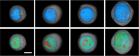
Three-dimensional images of a cell as it progresses from a normal cell to an invasive cancer. (Image from Dr. Deirdre Meldrum, Arizona State University)
Three-dimensional images of a cell as it progresses from a normal cell to an invasive cancer. Click to Enlarge (Image from Dr. Deirdre Meldrum, Arizona State University)

Researchers are now discovering what many have long suspected: much of what makes tumors heterogeneous overall is the substantial heterogeneity among individual cancer cells.

Until recently, the meticulous scrutiny of individual cells has been nearly impossible, particularly because of the relative scarcity in each cell of the key components that need to be measured, such as DNA and RNA. But thanks to technological advances that can help overcome some of those limitations, a growing number of investigators are beginning to delve deeper into the biology of the single cell.

The studies conducted to date “show us how much diversity there is among cancer cells in a given tumor,” said Dr. Garry Nolan, an immunologist at Stanford University whose lab is focused on mapping communication networks in individual cancer cells.

Even with improved technology, however, conducting studies at the single-cell level is difficult and can be time-consuming and expensive. But with growing interest—and $90 million over 5 years from the NIH Common Fund initiative (see the sidebar)—there is cautious optimism that over the next decade single-cell research may begin to pay dividends for patients with cancer and other diseases.

Moving beyond the Average

Most research on the molecular biology of tumors requires the use of mixtures of tens or hundreds of thousands of cells. Those samples “have immune cells, endothelial cells, and other infiltrating cells that make up the milieu of what a tumor actually is,” explained Dr. Dan Gallahan of NCI’s Division of Cancer Biology. “That really makes it difficult to get a grasp on what defines or, more importantly, how to treat a tumor.”

Results from studies that involve a bulk population of cells, Dr. Gallahan continued, essentially represent an average measurement.

Studying single cells is a way to “defy the average,” Dr. Marc Unger, chief scientific officer of Fluidigm Corporation, said earlier this year at a stem cell conference in Japan. (Fludigm, which develops tools for single-cell analysis, and the Broad Institute recently announced plans to establish a single-cell genomics research center.)

Single-cell analysis may be able to provide important clinical insights, said Dr. Nicholas Navin of the University of Texas MD Anderson Cancer Center, who has used next-generation sequencing to study variations in the number of genes (copy number variation) in single cancer cells.

Thanks to technological advances, a growing number of investigators are beginning to delve deeper into the biology of the single cell.

Single-cell analysis might, for example, help identify “pre-existing [cell populations] that are resistant to chemotherapy or rare subpopulations that are capable of invasion and metastasis,” he said. “We may also be able to quantify the extent of heterogeneity in a patient's tumor using single-cell data and use this index to predict how a patient will respond to treatment,” Dr. Navin continued.

Results from several recent studies have highlighted the challenges posed by tumor heterogeneity.

For example, researchers at BGI (formerly Beijing Genomics Institute) sequenced the protein-coding regions of DNA (the exome) of 20 cancer cells and 5 normal cells from a man with metastatic kidney cancer. The researchers found a tremendous amount of genetic diversity across the cancer cells, with very few sharing any common genetic mutations.

Much of the work in the analysis of single cells is still quite preliminary, and any potential clinical impact is still some years away, researchers agree.

“The problem with the single-cell data is that we don't really know yet what they mean,” Dr. Sangeeta Bhatia of the Massachusetts Institute of Technology commented recently in Nature Biotechnology.

And studies involving bulk populations of cells will not be going away any time soon, noted Dr. Betsy Wilder, director of the NIH Office of Strategic Coordination, which oversees the NIH Common Fund.

“Single-cell analysis isn’t warranted for every question that’s out there,” Dr .Wilder stressed. “Studies using populations of cells will continue to be done, because it makes a lot of sense to do them.”

Technology, a Driving Force

Beyond just an interest in learning more about single cells—what Dr. Gallahan called “the operational units in biology”—technology has been the driving force behind the growth of this field.

Dr. Stephen Quake, also of Stanford, has pioneered the use of microfluidics, which typically uses small chips with microscopic channels and valves—often called lab-on-a-chip devices—that allow researchers to single out and study individual cells. Dr. Quake, who co-founded Fluidigm, and others are increasingly using these devices for gene-expression profiling and for sequencing RNA and DNA of individual cells.

Dr. Nolan’s research involves a hybrid approach that combines two technologies: a souped-up method of flow cytometry, which has been used for several decades to sort cells and to perform limited analyses of single cells, and mass spectrometry, which is often used to identify and quantify proteins in biological samples.

Dr. Nolan’s lab is using this “mass cytometry” approach—developed by Dr. Scott Tanner of the University of Toronto—to characterize the response of individual cells to different stimuli, such as cytokines, growth factors, and a variety of drugs. Much of the group’s work has focused on analyzing normal blood-forming cells.

They published an influential study last year in Science that revealed some of the subtle biochemical changes that occur during cell differentiation. The study also described how dasatinib (Sprycel), a drug used to treat chronic myelogenous leukemia, affects certain intracellular activities. The research, Dr. Nolan said, is a prelude to studying individual cells from patients with blood cancers. The approach, he believes, may prove particularly useful for identifying new drugs and for testing them in the lab.

A tumor is not a uniform mass of identical cells with identical behaviors. Cells can act quite differently in one part of a tumor than in another.

The Microscale Life Sciences Center (MLSC), an NIH Center of Excellence in Genomic Science that is housed at Arizona State University, develops and applies the latest technology to single-cell research.

The center—a collaboration of investigators from Arizona State, the University of Washington, Brandeis University, and the Fred Hutchinson Cancer Research Center—includes researchers from numerous disciplines, including microfluidics, computer science, physics, engineering, and biochemistry, explained principal investigator Dr. Deirdre Meldrum.

“All of these disciplines are needed to develop the new technologies we’re working on,” said Dr. Meldrum, an electrical engineer by training.

In its initial work, the MLSC has measured metabolic processes in single living cells, including cellular respiration—the process by which cells acquire energy—as it relates to an individual cell’s ability to resist or succumb to cell death. The workhorse of this effort is a platform called the Cellarium, developed by Dr. Meldrum’s team. Individual cells are isolated in controlled chambers, Dr. Meldrum explained, “where we perturb them and measure how they change over time.”

Investigators at the MLSC and elsewhere have also developed technologies to image single cells. MLSC scientists are using a device developed by VisionGate, called the Cell-CT, “that enables accurate measurement of cellular features in true 3D,” Dr. Meldrum said.

MLSC researchers have studied abnormal esophageal cells from people with Barrett esophagus, a condition that increases the risk of esophageal adenocarcinoma. In particular, they’ve looked at how these cells respond to very low oxygen levels, or hypoxia.

Acid reflux, which can cause Barrett esophagus, can damage the esophagus “and lead to transient hypoxia in the epithelial lining of the esophagus,” explained Dr. Thomas Paulson, an investigator at Fred Hutchinson. In effect, he continued, the Cellarium system provides a snapshot of how this hypoxic environment selects for variants of cells that are able to survive and grow in it, providing insights into the factors that influence the evolution of cells from normal to cancerous.

Although Dr. Paulson’s work at MLSC is focused on Barrett esophagus, he believes the approach represents an excellent model system for studying cancer risk in general.

“I think our understanding of what constitutes risk is probably going to change as we understand the types of changes that occur at the single-cell level” that can transform a healthy cell into a cancerous cell, he said.

Deeper Dives Ahead

There’s a general acknowledgement in the field that single-cell analysis still has important limitations. Technological improvements are needed that can allow for the same type of molecular and structural “deep dives” that can be achieved by studying batches of cells. And powerful computer programs will be needed to help interpret the data from single-cell studies.

In addition, the research will eventually have to move beyond the confines of the mostly artificial environments in which single cells are now being tested, Dr. Gallahan noted. “As the technology gets better, we should be able to do more of this work in an in vivo setting.”

Although much more work is needed, the potential for what can be learned from studying single cells is quite large, Dr. Nolan believes.

“The fact that we’ve been able to make good decisions and learn as much as we have, even at the level of resolution [of cell populations], means that there’s something of even greater value to mine when you get to the level of the single cell,” he said.

Carmen Phillips

Transforming the Field of Single-Cell Research

This month, the National Institutes of Health will announce grant recipients for the NIH Common Fund’s single-cell analysis program.

The program, which includes three funding opportunities, “is largely a technology building program,” explained Dr. Wilder. The NIH Common Fund launched this program now because “there’s a sense that the technologies exist that can enable us to do the sort of analysis required to look at single cells in their native environment,” such as in a piece of excised tissue.

Although the focus is on technology, an important goal of the initiative is to support research that will “identify a few general principles of how single cells behave in a complex environment,” added Dr. Ravi Basavappa, the program director for the single-cell analysis program.

From the planning discussions, it was clear that the program should not limit the types of technology under consideration, Dr. Wilder commented. “Our analysis indicated that there are a lot of possibilities, so we left it up to the imaginations of the investigators to determine what technologies would be most transformative for the field as a whole.”

Featured Clinical Trial

Treating KSHV-Associated Multicentric Castleman Disease

Dr. Thomas Uldrick
Dr. Thomas Uldrick

Name of the Trial
Pilot Study of Tocilizumab in Patients with Symptomatic Kaposi Sarcoma Herpesvirus (KSHV)-Associated Multicentric Castleman Disease (NCI-11-C-0233). See the protocol summary.

Principal Investigator
Dr. Thomas Uldrick, NCI Center for Cancer Research

Why This Trial Is Important
Multicentric Castleman disease (MCD) is a group of rare diseases of the lymphatic system. One form is associated with infection by a virus called human herpesvirus 8. This virus is also associated with a type of cancer called Kaposi sarcoma, so it is sometimes known as Kaposi sarcoma herpesvirus (KSHV). Kaposi sarcoma and KSHV-associated MCD are much more common in people who are infected with the human immunodeficiency virus (HIV).

People with KSHV-associated MCD often have Kaposi sarcoma, and some treatments for KSHV-associated MCD can make Kaposi sarcoma worse. NCI researchers in the HIV and AIDS Malignancy Branch at NCI’s Center for Cancer Research are studying new approaches to treating KSHV-associated MCD and are trying to find better ways to manage it in patients who have both conditions.

People with KSHV-associated MCD may develop a variety of symptoms, such as low blood-cell counts (cytopenias), low levels of albumin (hypoalbuminemia), fever, fatigue, weight loss and muscle wasting (cachexia), fluid buildup (edema) in the legs and abdomen, and enlarged lymph nodes and spleen. Untreated, these inflammatory symptoms can be life threatening.

Currently, KSHV-associated MCD is usually managed with one of two treatments: the biological agent rituximab or antiviral therapy. Rituximab may improve symptoms, but it can also cause considerable side effects and lead to worsening Kaposi sarcoma. Virus-activated cytotoxic therapy, which was developed at NCI and consists of high-dose zidovudine (AZT) and valganciclovir, is one type of antiviral therapy that has shown some effectiveness against KSHV-associated MCD.

Although antiviral treatment targets the cause of the disease, it may not rapidly control the inflammatory symptoms. Therefore, doctors want to explore new approaches that may offer more effective and less-toxic treatment.

A monoclonal antibody called tocilizumab (Acterma) interferes with a molecular pathway that is important in MCD. The disease and many of its symptoms are driven by abnormal production of an inflammatory cytokine called interleukin-6 (IL-6). Tocilizumab attaches to the IL-6 receptor and prevents the molecular signaling that leads to IL-6-associated inflammation. This antibody was initially evaluated in the United States to treat rheumatoid arthritis and is approved by the Food and Drug Administration for that condition.

In this pilot study, patients with KSHV-associated MCD will receive intravenous tocilizumab every other week for up to 12 weeks. Patients who do not benefit from tocilizumab therapy alone may go on to receive high-dose AZT and valganciclovir in addition to tocilizumab. Doctors will assess the safety and effectiveness of tocilizumab alone and in combination with virus-activated cytotoxic therapy.

“Tocilizumab has been evaluated in other forms of MCD not related to KSHV and was effective in controlling the symptoms of the disease, including rapid resolution of hypoalbuminemia and anemia,” said Dr. Uldrick. “However, KSHV-associated MCD and other types of MCD are distinct diseases with overlapping clinical features. Tocilizumab may or may not be sufficient therapy in this setting, and that’s why we’re studying it, both alone and in combination with virus-activated cytotoxic therapy,” he said.

“The safety of tocilizumab is part of what we want to evaluate here; the major side effect we’d like to avoid is worsening Kaposi sarcoma in response to treating MCD,” Dr. Uldrick explained. “With rituximab, about half of the patients treated end up with worsening Kaposi sarcoma. That’s the yin-yang of treating these diseases; trying to manage KSHV-associated MCD without making Kaposi sarcoma worse.”

For More Information

See the lists of eligibility criteria and trial contact information or call the NCI Clinical Trials Referral Office at 1-888-NCI-1937. The call is toll free and confidential.

An archive of "Featured Clinical Trial" columns is available at http://www.cancer.gov/clinicaltrials/featured.
AHRQ Update

Task Force Reaffirms Recommendation against Ovarian Cancer Screening

Women at average risk of ovarian cancer should not be screened for the disease, the U.S. Preventive Services Task Force (USPSTF) has reaffirmed. Published in the Annals of Internal Medicine on September 11, the latest USPSTF clinical guideline does not apply to women who have symptoms of ovarian cancer or who have genetic mutations that increase their risk of ovarian cancer. Although available evidence does not show with absolute certainty whether the balance of benefits and harms of ovarian cancer screening differ for women with a family history of ovarian cancer, the USPSTF found no reason to believe that such women would necessarily benefit.

In 2008, the USPSTF commissioned the first review of the literature since its last recommendation on ovarian cancer screening in 2004. This review was extended through 2011 to consider the latest evidence from several randomized controlled trials, including results from the NCI-sponsored Prostate, Lung, Colorectal and Ovarian (PLCO) Cancer Screening Trial.

The review focused on two screening methods, transvaginal ultrasound and serum CA-125 testing, and found that screening not only failed to reduce ovarian cancer deaths but could also cause harm. For instance, the USPSTF noted, in the PLCO trial, "nearly 21 major complications occurred per 100 surgical procedures done on the basis of false-positive screening results." The USPSTF thus concluded that "there is at least moderate certainty that the harms of screening for ovarian cancer outweigh the benefits."

The USPSTF recommendation aligns with those of the American Cancer Society and the American Congress of Obstetricians and Gynecologists, neither of which recommend routine ovarian cancer screening for women of average risk without symptoms.

Further reading: "Contrary to Evidence, Some Doctors Recommend Ovarian Cancer Screening" and "Ovarian Cancer Screening Method Fails to Reduce Deaths from the Disease"

FDA Update

FDA Approves Imaging Agent that Helps Detect Prostate Cancer

The Food and Drug Administration (FDA) has approved the production and use of Choline C 11 Injection, an imaging agent used with positron emission tomography (PET) scans to help detect prostate cancer that has returned.

Choline C 11 Injection is administered intravenously to produce an image that helps to identify specific body sites for follow-up tissue sampling and testing in men whose prostate-specific antigen (PSA) levels have risen after earlier prostate cancer treatment.

The imaging agent must be made in a specialized facility and used shortly after its production. The Mayo Clinic is now the first FDA-approved facility to produce Choline C 11 Injection.

The safety and effectiveness of the imaging agent were verified by four independent studies. The studies examined a total of 98 patients with elevated PSA levels but no sign of recurrent prostate cancer with conventional imaging.

In each of the four studies, recurrent prostate cancer was confirmed by tissue sampling in at least half of the men who had abnormalities detected on PET scans with Choline C 11 imaging. PET scan errors were also reported. Depending on the study, false-positive PET scans were observed in 15 to 47 percent of the men. These findings underscore the need to confirm findings with tissue samples when abnormalities are detected with Choline C 11 Injection PET scans, the FDA noted in a press release.

Aside from a mild skin reaction at the injection site, no side effects to Choline C 11 Injection were reported.

Drug for Advanced Prostate Cancer Approved

The Food and Drug Administration (FDA) has approved enzalutamide (Xtandi) to treat men with advanced prostate cancer that has spread or recurred after medical or surgical therapy to minimize testosterone, which fuels tumor growth. The drug was approved for use in prostate cancer patients previously treated with docetaxel.

The safety and effectiveness of enzalutamide—previously called MDV3100—was evaluated in a study of 1,199 patients with metastatic castration-resistant prostate cancer who had received prior treatment with docetaxel. The median overall survival for patients who received enzalutamide was 18.4 months, compared with 13.6 months for those who received a placebo.

The most common side effects were fatigue, back pain, diarrhea, joint pain, hot flush, tissue swelling, musculoskeletal pain, respiratory infections, dizziness, spinal cord compression, blood in urine, tingling sensation, anxiety, and high blood pressure.

Seizures occurred in about 1 percent of those receiving enzalutamide. Study participants who had a seizure stopped enzalutamide therapy. The clinical study excluded men who had a history of seizure or several other brain conditions or who were taking medications that may cause seizures. The safety of enzalutamide in patients with these conditions is unknown.

Enzalutamide was reviewed under the FDA’s priority review program, which allows an expedited 6-month review for drugs that may offer major advances in treatment or that provide a treatment when no adequate therapy exists.

FDA Approves New Drug to Treat Chronic Myelogenous Leukemia

The Food and Drug Administration has approved bosutinib (Bosulif) to treat chronic myelogenous leukemia (CML), a blood and bone marrow disease that usually affects older adults. Bosutinib is intended for patients with chronic, accelerated, or blast phase Philadelphia chromosome-positive CML who are resistant to or who cannot tolerate other therapies, including imatinib (Gleevec).

Most people with CML have a chromosomal aberration called the Philadelphia chromosome, which causes the bone marrow to make an abnormal tyrosine kinase enzyme called Bcr-Abl. This enzyme promotes the proliferation of abnormal and unhealthy infection-fighting white blood cells called granulocytes. Bosutinib is a tyrosine kinase inhibitor (TKI) that works by blocking Bcr-Abl signaling.

Bosutinib’s safety and effectiveness were evaluated in a clinical trial involving 546 adults with chronic, accelerated, or blast phase CML. All of the patients had been previously treated with at least one TKI, either imatinib or imatinib followed by dasatinib (Sprycel) and/or nilotinib (Tasigna).

Among patients with chronic phase CML, 34 percent of patients who had been treated previously with imatinib and 27 percent of those who received more than one prior TKI achieved a major cytogenetic response within 24 weeks. 

Among patients with accelerated phase CML who had received at least one prior TKI, 30 percent had their blood counts return to the normal range (a complete hematologic response) by week 48, and 55 percent achieved a complete hematologic response, no evidence of leukemia, or return to chronic phase (an overall hematologic response) by week 48. Among patients with blast phase CML who had received at least one prior TKI, 15 percent had a complete hematologic response and 28 percent an overall hematologic response by week 48.

The most common side effects observed in those receiving bosutinib were diarrhea, nausea, a low level of platelets in the blood, vomiting, abdominal pain, rash, anemia, fever, and fatigue.

Notes

Cancer Classroom Webinar: Culture and Literacy

Cancer Classroom banner

As part of its Cancer Classroom Series, NCI is offering a webinar titled “Culture and Literacy: Bridging Communications Gaps” on September 27 from 2:00 p.m. to 3:00 p.m. ET.

Culture has been shown to influence people’s perceptions of cancer risk, their trust in oncology professionals, and their approach to cancer treatment including clinical research. Dr. Cathy Meade, director of the education program at the H. Lee Moffitt Cancer Center and Research Institute, will describe how incorporating culture into clinical practice may help reduce cancer health disparities across the cancer care continuum.

At the end of the presentation, participants will be able to

  • identify the role of culture and literacy in improving cancer communications;
  • examine the unique contributions of culture and literacy for reducing health disparities;
  • identify ways to integrate cultural and literacy considerations into community outreach programs, clinical practice, and innovations such as biobanking; and
  • identify resources relating to cancer, culture, and literacy.

The Cancer Classroom Series provides early career public health professionals and those new to oncology the educational guidance, tools, and resources necessary to address cancer as a public health issue.

Workshops are free, but preregistration is required and space is limited. To register for “Culture and Literacy: Bridging Communications Gaps” or to find out about upcoming sessions, visit the series website.

Call for Applications: NCI Clinical Assay Development Program

The NCI Clinical Assay Development Program (CADP) is requesting project applications from investigators seeking resources for clinical assay validation. These resources are designed to assist with the development of assays that may predict therapy response or prognostic behavior of a diagnosed cancer, primarily for use in clinical trials. Approved projects will have access to the institute’s assay development and validation resources, including project management support.

When applying to CADP, investigators must define the intended clinical use for the assay for which support is requested and provide basic assay protocols. Assays submitted for CADP development services should have been tested on human tissue. Proposals will be reviewed for scientific merit, feasibility, and clinical importance.

Applications are due October 15. More information and the application are available online.

New Brochure Educates Patients about Biospecimen Donation

Cover of How You Can Help Medical Research: Donating Your Blood, Tissue, and Other Samples

A new brochure, How You Can Help Medical Research: Donating Your Blood, Tissue, and Other Samples, explains how donating samples can help advance cancer research. The brochure, developed by NCI’s Office of Biorepositories and Biospecimen Research and Office of Communications and Education, covers common terms, explains the importance of donation, addresses potential concerns, and encourages readers to speak with their health care providers.

The brochure also provides answers to questions and concerns patients may have about biospecimen donation, helping readers make a more-informed decision about whether donation is right for them.

Readers may view the brochure and order a copy online.