
Unusual Cancers of the Reproductive and Urinary Systems
Bladder Cancer
Testicular Cancer
Ovarian Cancer
Cervical and Vaginal Cancer
Bladder Cancer
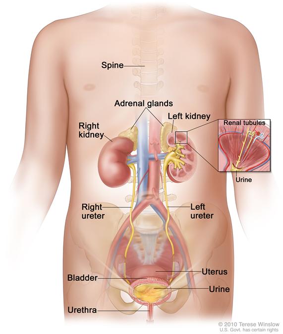
Bladder cancer is a disease in which malignant (cancer) cells form in the tissues of the bladder. The bladder is a hollow organ in the lower part of the abdomen. It is shaped like a small balloon and has a muscle wall that allows it to get bigger or smaller. The bladder stores urine until it is passed out of the body. Urine is the liquid waste that is made by the kidneys when they clean the blood. The urine passes from the two kidneys into the bladder through two tubes called ureters. When the bladder is emptied during urination, the urine goes from the bladder to the outside of the body through another tube called the urethra.
The most common type of bladder cancer is transitional cell cancer. Squamous cell and other more aggressive types of bladder cancer are less common.
Risk Factors, Symptoms, and Diagnostic and Staging Tests
In teenagers who were treated with certain anticancer drugs for leukemia, the risk of bladder cancer is increased.
Bladder cancer may cause any of the following signs and symptoms. Check with your doctor if any of the following occur:
- Blood in the urine (slightly rusty to bright red in color).
- Frequent urination or feeling the need to urinate without being able to do so.
- Pain during urination.
- Lower back pain.
Other conditions that are not bladder cancer may cause the same symptoms.
Tests to diagnose and stage bladder cancer may include the following:
- Physical exam and history.
- CT scan.
- Ultrasound of the bladder.
- Biopsy.
See the General Information section for a description of these tests and procedures.
Other tests used to diagnose bladder cancer include the following:
- Urinalysis: A test to check the color of urine and its contents, such as sugar, protein, red blood cells, and white blood cells.
- Urine cytology: Examination of urine under a microscope to check for abnormal cells.
- Cystoscopy: A procedure to look inside the bladder and urethra to check for abnormal areas. A cystoscope is inserted through the urethra into the bladder. A cystoscope is a thin, tube-like instrument with a light and a lens for viewing. It may also have a tool to remove tissue samples, which are checked under a microscope for signs of cancer.
Prognosis
In children, bladder cancer is usually low grade (not likely to spread) and the prognosis is usually good following surgery to remove the tumor.
Treatment
Treatment for bladder cancer in children is usually transurethral resection (TUR). This is a surgical procedure to remove tissue from the bladder using a resectoscope inserted into the bladder through the urethra. A resectoscope is a thin, tube-like instrument with a light, a lens for viewing, and a tool to remove tissue and burn away any remaining tumor cells. Tissue samples are checked under a microscope for signs of cancer.
See the PDQ summary on adult Bladder Cancer Treatment for more information.
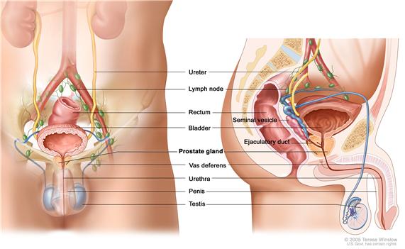
Testicular CancerTesticular cancer is a disease in which malignant (cancer) cells form in the tissues of one or both testicles. The testicles are 2 egg-shaped glands located inside the scrotum (a sac of loose skin that lies directly below the penis). The testicles are held within the scrotum by the spermatic cord, which also contains the vas deferens and vessels and nerves of the testicles.
There are two types of testicular tumors:
- Germ cell tumors: Tumors that start in sperm cells in males. Testicular germ cell tumors may be benign (not cancer) or malignant (cancer). The most common testicular germ cell tumors in young boys are benign teratomas and malignant nonseminomas. Seminomas usually occur in young men and are rare in boys.
- Non-germ cell tumors: Tumors that begin in the tissues that surround and support the testicles. These tumors may be benign or malignant.
Symptoms and Diagnostic and Staging Tests
A testicular tumor may cause a painless lump in the testicles. Other conditions may also cause a lump in the testicles.
Tests to diagnose and stage non-germ cell testicular cancer may include the following:
See the General Information section for a description of these tests and procedures.
Treatment
Treatment for non-germ cell testicular cancer in children may be surgery.
See the PDQ summary on Childhood Extracranial Germ Cell Tumors Treatment for more information on testicular germ cell tumors.
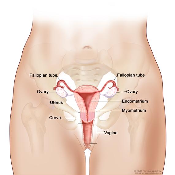
Ovarian CancerOvarian cancer is a disease in which malignant (cancer) cells form in the ovary. The ovaries are a pair of organs in the female reproductive system. They are located in the pelvis, one on each side of the uterus (the hollow, pear-shaped organ where a fetus grows). Each ovary is about the size and shape of an almond. The ovaries produce eggs and female hormones (chemicals that control the way certain cells or organs function).
Most ovarian tumors in children are benign (not cancer). They occur most often in females aged 15 to 19 years.
There are several common types of malignant ovarian tumors:
- Germ cell tumors: Tumors that start in egg cells in females. These are the most common ovarian tumors in girls. (See the PDQ summary on Childhood Extracranial Germ Cell Tumors Treatment for more information on ovarian germ cell tumors.)
- Epithelial tumors: Tumors that start in the tissue covering the ovary. These are the second most common ovarian tumors in girls.
- Stromal tumors: Tumors that begin in stromal cells, which make up tissues that surround and support the ovaries.
- Other tumors, such as Burkitt lymphoma and small cell carcinoma of the ovary (a very rare tumor).
Risk Factors, Symptoms, and Diagnostic and Staging Tests
The risk of ovarian cancer is increased by having one of the following conditions:
- Ollier disease (a disorder that causes abnormal growth of cartilage at the end of long bones).
- Maffucci syndrome (a disorder that causes abnormal growth of cartilage at the end of long bones and of blood vessels in the skin).
- Peutz-Jeghers syndrome.
Ovarian cancer may cause any of the following signs and symptoms. Check with your doctor if any of the following problems occur:
- Painful menstrual periods.
- A lump in the abdomen.
- Pain or swelling in the abdomen.
- Having male sex traits, such as body hair or a deep voice.
- Early signs of puberty.
Other conditions that are not ovarian cancer may cause these same symptoms.
Tests to diagnose and stage ovarian cancer may include the following:
See the General Information section for a description of these tests and procedures.
Prognosis
Ovarian epithelial cancer is usually found at an early stage in children and is easier to treat than in adult patients.
Treatment
Treatment of ovarian epithelial cancer may include the following:
Treatment of ovarian stromal tumors may include the following:
- Surgery to remove one ovary and one fallopian tube, for early cancer.
- Surgery followed by chemotherapy for cancer that is advanced.
- Chemotherapy for cancer that has recurred (come back).
See the following PDQ summaries for more information:
- Childhood Extracranial Germ Cell Tumors Treatment
- Ovarian Epithelial Cancer Treatment
- Ovarian Germ Cell Tumors Treatment
Cervical cancer is a disease in which malignant (cancer) cells form in the cervix. The cervix is the lower, narrow end of the uterus (the hollow, pear-shaped organ where a fetus grows). The cervix leads from the uterus to the vagina (birth canal). Vaginal cancer forms in the vagina. The vagina is the canal leading from the cervix to the outside of the body. At birth, a baby passes out of the body through the vagina (also called the birth canal).
The most common symptom of cervical and vaginal cancer is bleeding from the vagina. Other conditions may also cause vaginal bleeding.
Treatment
Treatment for childhood cervical and vaginal cancer may include surgery to remove as much of the cancer as possible, followed by radiation therapy. Chemotherapy may also be used but it is not yet known if this is an effective treatment.
 Back to Top