
Rabies and Kids!
Immunohistochemistry (IHC)
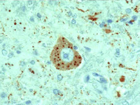
This slide shows a rabies-infected neuronal cell with intracytoplasmic inclusions. The red stain indicates areas of rabies viral antigen by using IHC or avidin-biotin complex (ABC) technique.
IHC methods for rabies detection provide sensitive and specific means to detect rabies in formalin-fixed tissues. These methods are more sensitive than histologic staining methods, such as H&E and Sellers stains. Like the dFA test, these procedures use specific antibodies to detect rabies virus inclusions. The techniques use enzyme-labeling systems that increase sensitivity. In addition, monoclonal antibodies may be used to detect rabies virus variants.
Get email updates
To receive email updates about this page, enter your email address:
Contact Us:
- Centers for Disease Control and Prevention
1600 Clifton Rd
Atlanta, GA 30333 - 800-CDC-INFO
(800-232-4636)
TTY: (888) 232-6348 - New Hours of Operation
8am-8pm ET/Monday-Friday
Closed Holidays - cdcinfo@cdc.gov