


How the Lungs Work
To understand idiopathic pulmonary fibrosis (IPF), it helps to understand how the lungs work. The air that you breathe in through your nose or mouth travels down through your trachea (windpipe) into two tubes in your lungs called bronchial (BRONG-ke-al) tubes or airways.
The airways are shaped like an upside-down tree with many branches. The windpipe is the trunk. It splits into two bronchial tubes, or bronchi. Thinner tubes called bronchioles branch out from the bronchi.
The bronchioles end in tiny air sacs called alveoli (al-VEE-uhl-eye). These air sacs have very thin walls, and small blood vessels called capillaries run through them. There are about 300 million alveoli in a normal lung.
When the air that you've just breathed in reaches these air sacs, the oxygen in the air passes through the air sac walls into the blood in the capillaries. At the same time, carbon dioxide (a waste gas) moves from the capillaries into the air sacs. This process is called gas exchange.
The oxygen-rich blood in the capillaries then flows into larger veins, which carry it to the heart. Your heart pumps the oxygen-rich blood to all your body's organs. These organs can't function without an ongoing supply of oxygen.
The animation below shows how the lungs work. Click the "start" button to play the animation. Written and spoken explanations are provided with each frame. Use the buttons in the lower right corner to pause, restart, or replay the animation, or use the scroll bar below the buttons to move through the frames.
The animation shows how the lungs inhale oxygen and transfer it to the blood. It also shows how carbon dioxide (a waste product) is removed from the blood and exhaled.
In IPF, scarring begins in the air sac walls and the spaces around them. The scarring makes the walls of the air sacs thicker. This makes it harder for oxygen to pass through the air sac walls into the bloodstream.
Idiopathic Pulmonary Fibrosis
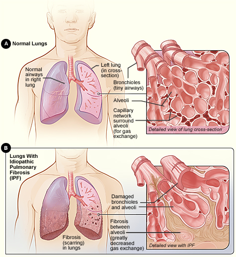
Figure A shows the location of the lungs and airways in the body. The inset image shows a detailed view of the lung's airways and air sacs in cross-section. Figure B shows fibrosis (scarring) in the lungs. The inset image shows a detailed view of the fibrosis and how it damages the airways and air sacs.
For more information about lung function, go to the Health Topics How the Lungs Work article.


Clinical trials are research studies that explore whether a medical strategy, treatment, or device is safe and effective for humans. To find clinical trials that are currently underway for Idiopathic Pulmonary Fibrosis, visit www.clinicaltrials.gov.
October 21, 2011
Commonly used three-drug regimen for idiopathic pulmonary fibrosis found harmful
The National Heart, Lung, and Blood Institute (NHLBI), part of the National Institutes of Health, has stopped one arm of a three arm multi-center, clinical trial studying treatments for the lung-scarring disease idiopathic pulmonary fibrosis (IPF) for safety concerns.
The NHLBI updates Health Topics articles on a biennial cycle based on a thorough review of research findings and new literature. The articles also are updated as needed if important new research is published. The date on each Health Topics article reflects when the content was originally posted or last revised.