Useful Links
Newsroom Image Library
Disease Agents
These images are in the public domain and are thus free of any copyright restrictions. As a matter of courtesy, we request that the content provider be credited and notified of any public or private usage of an image.
For more information on an image, including the content provider(s) and extended descriptions, please enter the image’s PHIL ID number in the search box in the Public Health Image Library. If you need assistance, please e-mail media@cdc.gov or call 404-639-3286.
2009 H1N1 Flu

PHIL ID #11822
Photo Credit: Illustrator: Dan Higgins, CDC
Download High Resolution
2009 H1N1 Flu
PHIL ID #11823
Photo Credit: Illustrator: Dan Higgins, CDC
Download High Resolution
2009 H1N1 Flu

PHIL ID #11215
Photo Credit: C. S. Goldsmith and A. Balish, CDC
Download High Resolution
2009 H1N1 Flu

PHIL ID #11214
Photo Credit: C. S. Goldsmith and A. Balish, CDC
Download High Resolution
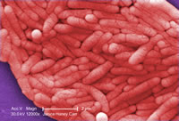
Legionella (Legionnaire′s Disease)

PHIL ID #9999
Photo Credit: Janice Haney Carr, Centers for Disease Control and Prevention
Download High Resolution
Clostridium difficile

PHIL ID #9999
Photo Credit: Janice Haney Carr, Centers for Disease Control and Prevention
Download High Resolution
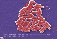
Salmonella typhimurium
PHIL ID #10983
Photo Credit: Janice Haney Carr, Centers for Disease Control and Prevention
Download High Resolution
Salmonella typhimurium

PHIL ID #10971
Photo Credit: Janice Haney Carr, Centers for Disease Control and Prevention
Download High Resolution
Salmonella

PHIL ID #10896
Photo Credit: Janice Haney Carr, Centers for Disease Control and Prevention
Download High Resolution
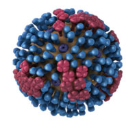
Influenza virus particle

PHIL ID #10073
Photo Credit: Cynthia Goldsmith, Centers for Disease Control and Prevention
Download High Resolution
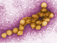
West Nile Virus

PHIL ID #10700
Photo Credit: Cynthia Goldsmith, Centers for Disease Control and Prevention
Download High Resolution
West Nile Virus
PHIL ID #10701
Photo Credit: Cynthia Goldsmith, Centers for Disease Control and Prevention
Download High Resolution
Group C Streptococcus
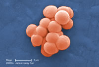
PHIL ID #10586
Photo Credit: Janice Haney Carr, Centers for Disease Control and Prevention
Download High Resolution
Group C Streptococcus

PHIL ID #10591
Photo Credit: Janice Haney Carr, Centers for Disease Control and Prevention
Download High Resolution
Measles

PHIL ID #10707
Photo Credit: Cynthia Goldsmith, Centers for Disease Control and Prevention
Download High Resolution
Norovirus
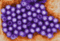
PHIL ID #10708
Photo Credit: Charles D. Humphrey, Centers for Disease Control and Prevention
Download High Resolution
Norovirus

PHIL ID #10709
Photo Credit: Charles D. Humphrey, Centers for Disease Control and Prevention
Download High Resolution
Avian Influenza A H5N1

PHIL ID # 1841
Photo Credit: Cynthia Goldsmith, Centers for Disease Control and Prevention
Download High Resolution
Avian influenza A viruses do not usually infect humans; however, several instances of human infections and outbreaks have been reported since 1997. When such infections occur, public health authorities monitor these situations closely.
MRSA

PHIL ID # 10045
Photo Credit: Janice Carr, Centers for Disease Control and Prevention
Download High Resolution
MRSA

PHIL ID # 10046
Photo Credit: Janice Haney, Centers for Disease Control and Prevention
Download High Resolution
E. Coli
PHIL ID # 10068
Photo Credit: Janice Haney Carr, Centers for Disease Control and Prevention
Download High Resolution
Tuberculosis
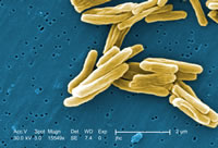
PHIL ID # 9997
Photo Credit: Janice Carr, Centers for Disease Control and Prevention
Download High Resolution
HIV-1

PHIL ID # 10000
Photo Credit: Cynthia Goldsmith, Centers for Disease Control and Prevention
Download High Resolution
- Page last reviewed: October 15, 2009
- Page last updated: October 15, 2009
- Content source: Office of the Associate Director for Communication
- Notice: Links to non-governmental sites do not necessarily represent the views of the CDC.
View Press Releases in

Contact Us:
- Centers for Disease Control and Prevention
1600 Clifton Rd
Atlanta, GA 30333 - 800-CDC-INFO
(800-232-4636)
TTY: (888) 232-6348 - Contact CDC-INFO