
Unusual Cancers of the Chest
Breast Cancer
Lung Cancer
Bronchial Tumors
Pleuropulmonary Blastoma
Esophageal Tumors
Thymoma and Thymic Carcinoma
Heart Tumors
Mesothelioma
Breast Cancer
Most breast tumors in children are fibroadenomas, which are benign (not cancer). Rarely, these tumors become large phyllodes tumors (cancer) and begin to grow quickly. If a benign tumor begins to grow quickly, a fine-needle aspiration (FNA) biopsy or an excisional biopsy will be done. The tissues removed during the biopsy will be viewed under a microscope by a pathologist to check for signs of cancer.
Breast cancer is a disease in which malignant (cancer) cells form in the tissues of the breast. Breast cancer may occur in both male and female children.
Breast cancer is the most common cancer among teenage and young adult women aged 15 to 39 years. Breast cancer in this age group is more aggressive and more difficult to treat successfully than in older women. Treatments for younger and older women are similar. Also, care for younger patients with breast cancer includes checking for familial cancer syndromes and considering possible fertility issues when choosing treatment.
Risk Factors, Symptoms, and Diagnostic and Staging Tests
The risk of breast cancer is increased by the following:
- Having a personal history of cancer that may spread to the breast, such as leukemia, rhabdomyosarcoma, soft tissue sarcoma, or lymphoma.
- Past treatment for another cancer, such as Hodgkin lymphoma, with radiation therapy to the breast or chest.
Breast cancer may cause any of the following signs and symptoms. Check with your doctor if any of the following problems occur:
- A lump or thickening in or near the breast or in the underarm area.
- A change in the size or shape of the breast.
- A dimple or puckering in the skin of the breast.
- A nipple turned inward into the breast.
- Scaly, red, or swollen skin on the breast, nipple, or areola (the dark area of skin that is around the nipple).
- Dimples in the breast that look like the skin of an orange, called peau d’orange.
Other conditions that are not breast cancer may cause these same symptoms.
Tests to diagnose and stage breast cancer may include the following:
- Physical exam and history.
- MRI.
- Ultrasound.
- PET scan.
- Blood chemistry studies.
- X-ray of the chest.
- Biopsy.
See the General Information section for a description of these tests and procedures.
Another test used to diagnose breast cancer is the mammogram (an x-ray of the breast). When treatment for another cancer included radiation therapy to the breast or chest, it is important to have a mammogram and MRI of the breast to check for breast cancer beginning at age 25, or 10 years after finishing radiation therapy, whichever is later.
Treatment
Treatment of breast cancer in children may include the following:
- Watchful waiting, for benign tumors.
- Surgery to remove the tumor, but not the whole breast. Radiation therapy may also be given.
See the PDQ summary Breast Cancer Treatment for more information on the treatment of adolescents and young adults with breast cancer.
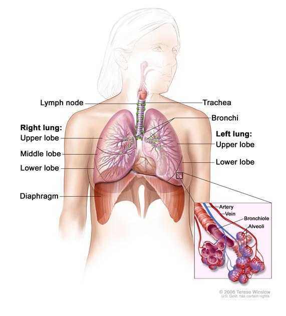
Lung CancerLung cancer begins in the tissue of the lung. The lungs are a pair of cone-shaped breathing organs in the chest. The lungs bring oxygen into the body as you breathe in. They release carbon dioxide, a waste product of the body’s cells, as you breathe out. Each lung has sections called lobes. The left lung has two lobes. The right lung is slightly larger and has three lobes. Two tubes called bronchi lead from the trachea (windpipe) to the right and left lungs. Tiny air sacs called alveoli and small tubes called bronchioles make up the inside of the lungs.
In children, most lung tumors are malignant (cancer).
Symptoms and Diagnostic Tests
Lung cancer may cause any of the following signs and symptoms. Check with your doctor if any of the following problems occur:
- Coughing.
- Streaks of blood in sputum (mucus coughed up from the lungs).
- Trouble breathing.
- Chest discomfort.
- Fever.
- Weight loss for no known reason.
Tests to diagnose lung cancer may include the following:
- Physical exam and history.
- X-ray of the chest.
- CT scan.
See the General Information section for a description of these tests and procedures.
Treatment
Treatment for lung cancer in children is surgery to remove the tumor. More treatment may be given after surgery. It depends on the type of tumor and whether the tumor has spread.
Bronchial TumorsBronchial tumors begin in the cells that line the surface of the lung. Most bronchial tumors in children are benign, slow-growing tumors in the trachea or large bronchi, which are the large airways of the lung. Sometimes, a slow-growing bronchial tumor becomes cancer that may spread to other parts of the body.
Symptoms and Diagnostic and Staging Tests
Bronchial tumors may cause any of the following signs and symptoms:
- Coughing.
- Wheezing.
- Trouble breathing.
- Spitting up blood from the airways or lung.
- Frequent infections in the lung, such as pneumonia.
Other conditions that are not bronchial tumors may cause these same symptoms. For example, symptoms of bronchial tumors are a lot like the symptoms of asthma, and that can make it hard to diagnose the tumor.
Tests to diagnose and stage bronchial tumors may include the following:
- Physical exam and history.
- X-ray of the chest.
- CT scan.
See the General Information section for a description of these tests and procedures.
A biopsy of the abnormal area is usually not done because it can cause severe bleeding.
Other tests used to diagnose bronchial tumors include the following:
- Bronchoscopy: A procedure to look inside the trachea and large airways in the lung for abnormal areas. A bronchoscope is inserted through the nose or mouth into the trachea and lungs. A bronchoscope is a thin, tube-like instrument with a light and a lens for viewing. It may also have a tool to remove tissue samples, which are checked under a microscope for signs of cancer. A contrast dye may be put through the bronchoscope to make the larynx, trachea, and airways show up clearer on x-ray film.
- Octreotide scan: A type of radionuclide scan used to find tumors. A small amount of radioactive octreotide (a hormone that attaches to carcinoid tumors) is injected into a vein and travels through the bloodstream. The radioactive octreotide attaches to the tumor and a special camera that detects radioactivity is used to show where the tumors are in the body.
Prognosis
Bronchial cancer in children can usually be cured, even when it has spread to nearby areas. The prognosis (chance of recovery) depends on how the cells look under a microscope and the stage of the cancer.
Treatment
Treatment of bronchial tumors in children may include the following:
- Surgery to remove the tumor. Sometimes a type of surgery called a sleeve resection is used. The lymph nodes and vessels where cancer has spread are also removed.
- Chemotherapy or radiation therapy, for cancer that has spread to other parts of the body.
Pleuropulmonary blastomas (PPBs) form in the tissue of the lung and pleura (tissue that covers the lungs and lines the inside of the chest). PPBs can also form in the organs between the lungs including the heart, aorta, and pulmonary artery, or in the diaphragm (the main breathing muscle below the lungs).
There are three stages of PPB that are described as types:
- Type I tumors are cyst -like tumors in the lung. They are most common in children aged 2 years and younger and can usually be cured.
- Type II tumors are cyst-like with some solid parts. These tumors sometimes spread to the brain.
- Type III tumors are solid. These tumors often spread to the brain.
Risk Factors, Symptoms, and Diagnostic and Staging Tests
The risk of PPB is increased by the following:
- Having a family history of any type of cancer in close relatives.
- Having a brother or sister with PPB.
- Having a personal history of other types of cancer.
PPB may cause any of the following signs and symptoms. Check with your doctor if any of the following problems occur:
- A cough that doesn’t go away.
- Trouble breathing.
- Chest discomfort.
- Wheezing.
- Streaks of blood in sputum (mucus coughed up from the lungs).
- Hoarseness.
- Pain under the rib cage.
- Pain, swelling, or lumps in the abdomen.
- Loss of appetite.
- Weight loss for no known reason.
- Feeling very tired.
Other conditions that are not PPB may cause these same symptoms.
Tests to diagnose and stage PPB may include the following:
- Physical exam and history.
- X-ray of the chest.
- CT scan.
- PET scan.
See the General Information section for a description of these tests and procedures.
Other tests used to diagnose PPB include the following:
- Bronchoscopy: A procedure to look inside the trachea and large airways in the lung for abnormal areas. A bronchoscope is inserted through the nose or mouth into the trachea and lungs. A bronchoscope is a thin, tube-like instrument with a light and a lens for viewing. It may also have a tool to remove tissue samples, which are checked under a microscope for signs of cancer.
- Thoracoscopy: A surgical procedure to look at the organs inside the chest to check for abnormal areas. An incision (cut) is made between two ribs, and a thoracoscope is inserted into the chest. A thoracoscope is a thin, tube-like instrument with a light and a lens for viewing. It may also have a tool to remove tissue or lymph node samples, which are checked under a microscope for signs of cancer. In some cases, this procedure is used to remove part of the esophagus or lung. If the thoracoscope cannot reach certain tissues, organs, or lymph nodes, a thoracotomy may be done. In this procedure, a larger incision is made between the ribs and the chest is opened.
PPBs may spread or recur (come back) even after being removed by surgery.
Treatment
Treatment of pleuropulmonary blastomas in children is usually surgery to remove the whole lobe of the lung the tumor is in, with or without chemotherapy.
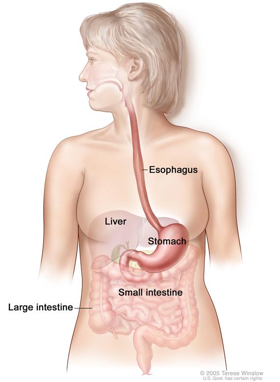
Esophageal TumorsEsophageal tumors may be benign (not cancer) or malignant (cancer). Esophageal cancer is a disease in which malignant cells form in the tissues of the esophagus. The esophagus is the hollow, muscular tube that moves food and liquid from the throat to the stomach. Most esophageal tumors in children begin in the thin, flat cells that line the esophagus.
Symptoms and Diagnostic and Staging Tests
Esophageal cancer may cause any of the following signs and symptoms. Check with your doctor if any of the following problems occur:
- Trouble swallowing.
- Weight loss.
- Pain behind the breastbone.
- Hoarseness and cough.
- Indigestion and heartburn.
Other conditions that are not esophageal cancer may cause these same symptoms.
Tests to diagnose and stage esophageal cancer may include the following:
- Physical exam and history.
- X-ray of the chest.
- CT scan.
- PET scan.
- Ultrasound.
- Biopsy.
See the General Information section for a description of these tests and procedures.
Other tests used to diagnose esophageal cancer include the following:
- Esophagoscopy: A procedure to look inside the esophagus to check for abnormal areas. An esophagoscope is inserted through the mouth or nose and down the throat into the esophagus. An esophagoscope is a thin, tube-like instrument with a light and a lens for viewing. It may also have a tool to remove tissue samples, which are checked under a microscope for signs of cancer. A biopsy is usually done during an esophagoscopy. Sometimes a biopsy shows changes in the esophagus that are not cancer but may lead to cancer.
- Bronchoscopy: A procedure to look inside the trachea and large airways in the lung for abnormal areas. A bronchoscope is inserted through the nose or mouth into the trachea and lungs. A bronchoscope is a thin, tube-like instrument with a light and a lens for viewing. It may also have a tool to remove tissue samples, which are checked under a microscope for signs of cancer.
- Thoracoscopy: A surgical procedure to look at the organs inside the chest to check for abnormal areas. An incision (cut) is made between two ribs and a thoracoscope is inserted into the chest. A thoracoscope is a thin, tube-like instrument with a light and a lens for viewing. It may also have a tool to remove tissue or lymph node samples, which are checked under a microscope for signs of cancer. Sometimes this procedure is used to remove part of the esophagus or lung.
- Laparoscopy: A surgical procedure to look at the organs inside the abdomen to check for signs of disease. Small incisions (cuts) are made in the wall of the abdomen and a laparoscope (a thin, lighted tube) is inserted into one of the incisions. Other instruments may be inserted through the same or other incisions to perform procedures such as removing organs or taking tissue samples to be checked under a microscope for signs of disease.
Prognosis
Esophageal cancer is hard to cure because it usually is not possible to remove the whole tumor by surgery.
Treatment
Treatment for esophageal cancer in children may include the following:
- Surgery to remove all or part of the tumor.
- Radiation therapy given through a plastic or metal tube placed through the mouth into the esophagus.
- Chemotherapy.
See the PDQ summary on adult Esophageal Cancer for more information.
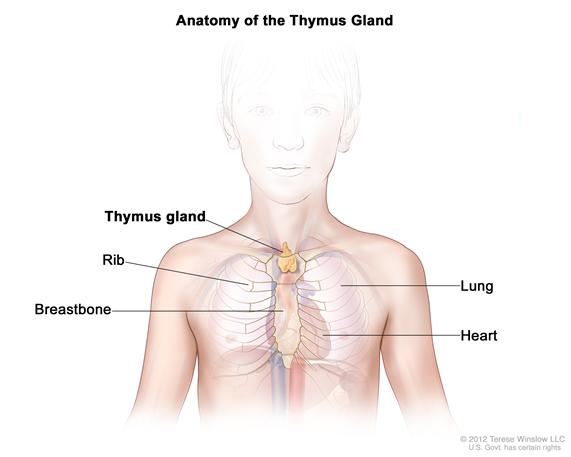
Thymoma and Thymic CarcinomaThymomas and thymic carcinomas are tumors of the cells that cover the outside surface of the thymus. The thymus is a small organ in the upper chest under the breastbone. It is part of the lymph system and makes white blood cells, called lymphocytes, that help fight infection. Thymomas and thymic carcinomas usually form in the front part of the chest and are often found during a chest x-ray that is done for another reason.
Thymoma and thymic carcinoma are slow-growing cancers that may spread to the lymph nodes or to other parts of the body.
Risk Factors, Symptoms, and Diagnostic and Staging Tests
People who develop thymomas often have one of the following immune system diseases or hormone disorders:
- Myasthenia gravis.
- Polymyositis.
- Lupus.
- Rheumatoid arthritis.
- Thyroiditis.
- Isaac syndrome.
- Pure red cell aplasia.
- Hyperthyroidism.
- Addison disease.
- Panhypopituitarism.
Thymoma and thymic carcinoma may cause any of the following symptoms Check with your doctor if any of the following problems occur:
- Coughing.
- Trouble swallowing.
- Pain or a tight feeling in the chest.
- Trouble breathing.
Other conditions that are not thymoma and thymic carcinoma may cause these same symptoms.
Tests to diagnose and stage thymoma and thymic carcinoma may include the following:
See the General Information section for a description of these tests and procedures.
Prognosis
The prognosis (chance of recovery) is better when the tumor has not spread.
Treatment
Treatment for thymomas and thymic carcinoma in children may include the following:
- Surgery to remove as much of the tumor as possible, followed by radiation therapy for tumors that have spread.
- Chemotherapy.
Most tumors that form in the heart are benign (not cancer). Benign heart tumors that may appear in children include the following:
- Rhabdomyoma: A tumor that forms in muscle made up of long fibers.
- Fibroma: A tumor that forms in fiber-like tissue that holds bones, muscles, and other organs in place.
- Myxoma: A tumor that may be part of an inherited syndrome called Carney complex. (See the Multiple Endocrine Neoplasia Syndromes section for more information.)
- Histiocytoid cardiomyopathy tumor: A tumor that forms in the heart cells that control heart rhythm.
- Teratomas: A type of germ cell tumor. In the heart, these tumors form most often in the pericardium (the sac that covers the heart). Some teratomas are malignant (cancer).
- Hemangiomas: A tumor that forms in the cells that line blood vessels.
- Neurofibroma: A tumor that forms in the cells and tissues that cover nerves.
In children, the most common benign heart tumors are rhabdomyomas and fibromas. Before birth and in newborns, the most common benign heart tumors are teratomas. An inherited disorder called tuberous sclerosis can cause heart tumors to form in a fetus or newborn.
Malignant tumors that begin in the heart are even more rare than benign tumors in children. Some of these include:
- Malignant teratoma.
- Rhabdomyosarcoma: A cancer that forms in muscle made up of long fibers.
- Chondrosarcoma: A type of cancer that usually forms in bone cartilage but very rarely can begin in the heart.
- Infantile fibrosarcoma.
Some cancers, such as rhabdomyosarcoma, melanoma, and leukemia, spread to the heart from other parts of the body. These tumors are malignant.
Symptoms
Heart tumors may cause any of the following symptoms. Check with your doctor if any of the following problems occur:
- Change in the heart's normal rhythm.
- Trouble breathing, particularly when you are lying down.
- Pain in the middle of the chest that feels better when you are sitting up.
- Coughing.
- Fainting.
- Feeling dizzy, tired, or weak.
- Fast heart rate.
- Swelling in the legs, ankles, or abdomen.
- Feeling anxious.
Heart tumors sometimes cause sudden death without causing any symptoms.
Other conditions that are not heart tumors may cause these same symptoms. Sometimes heart tumors do not cause any symptoms at all.
Tests to diagnose and stage heart tumors may include the following:
- Physical exam and history.
- X-ray of the chest.
- CT scan.
- MRI.
See the General Information section for a description of these tests and procedures.
Other tests used to diagnose or stage heart tumors include the following:
- Echocardiogram: A procedure in which high-energy sound waves (ultrasound) are bounced off the heart and nearby tissues or organs and make echoes. A moving picture is made of the heart and heart valves as blood is pumped through the heart.
- Electrocardiogram (EKG): A recording of the heart's electrical activity to evaluate its rate and rhythm. A number of small pads (electrodes) are placed on the patient’s chest, arms, and legs, and are connected by wires to the EKG machine. Heart activity is then recorded as a line graph on paper. Electrical activity that is faster or slower than normal may be a sign of heart disease or damage.
Treatment
Treatment for heart tumors in children may include the following:
- Watchful waiting for benign tumors of heart muscle (rhabdomyomas), which usually shrink and go away on their own.
- Surgery (which may include a heart transplant) and chemotherapy for tumors that spread to the heart from other places in the body.
Malignant mesothelioma is a disease in which malignant (cancer) cells are found in the pleura (the thin layer of tissue that lines the chest cavity and covers the lungs) or the peritoneum (the thin layer of tissue that lines the abdomen and covers most of the organs in the abdomen). The tumors often spread over the surface of organs without spreading into the organ. They may spread to lymph nodes nearby or in other parts of the body.
Risk Factors, Symptoms, and Diagnostic and Staging Tests
Mesothelioma is sometimes a late effect of treatment for an earlier cancer, especially after treatment with radiation therapy. In adults, mesothelioma has been linked to being exposed to asbestos, which was once used as building insulation. There is no information about the risk of mesothelioma in children exposed to asbestos.
Mesothelioma may cause any of the following signs and symptoms. Check with your doctor if any of the following problems occur:
- Trouble breathing.
- Pain under the rib cage.
- Weight loss for no known reason.
Other conditions that are not mesothelioma may cause these same symptoms.
Tests to diagnose and stage mesothelioma may include the following:
- Physical exam and history.
- X-ray of the chest.
- CT scan.
- PET scan.
- Fine-needle aspiration (FNA) biopsy.
See the General Information section for a description of these tests and procedures.
Other tests used to diagnose mesothelioma include the following:
- Bronchoscopy: A procedure to look inside the trachea and large airways in the lung for abnormal areas. A bronchoscope is inserted through the nose or mouth into the trachea and lungs. A bronchoscope is a thin, tube-like instrument with a light and a lens for viewing. It may also have a tool to remove tissue samples, which are checked under a microscope for signs of cancer.
- Thoracoscopy: An incision (cut) is made between two ribs and a thoracoscope (a thin, tube-like instrument with a light and a lens for viewing) is inserted into the chest to check for signs of disease.
- Thoracotomy: An incision (cut) is made between two ribs to check inside the chest for signs of disease.
- Cytologic exam: An exam of cells under a microscope (by a pathologist) to check for anything abnormal. For mesothelioma, fluid is taken from around the lungs or from the abdomen. A pathologist checks the cells in the fluid.
Prognosis
The prognosis (chance of recovery) is better when the tumor has not spread or come back after treatment.
Treatment
Treatment for mesothelioma in children may include one or more of the following:
- Surgery to remove the part of the chest lining with cancer and some of the healthy tissue around it.
- Chemotherapy.
- Radiation therapy, as palliative therapy, to relieve pain and improve quality of life.
See the PDQ summary on adult Malignant Mesothelioma Treatment for more information.
 Back to Top