


Genomics in Action: William A. Gahl, M.D., Ph.D.
Powerful Insights From Rare Problems
 What occurs in less than one-tenth of one percent of the population, yet is found in one form or another in almost every family in the world?
A rare genetic disease.
According to the Orphan Drug Act of 1983, a rare disease is defined as a disorder that occurs in less than 200,000 people in the United States, but many rare genetic diseases are substantially less common than this. For instance, National Human Genome Research Institute (NHGRI) Clinical Director William A. Gahl, M.D., Ph.D., has seen only five people with Chediak-Higashi disease over the course of his entire career. That's still more Chediak-Higashi patients than anyone else in the United States is currently treating.
Why does Dr. Gahl work with these patients? Couldn't his time be better spent on disorders that affect far more people or have a chance of being cured?
"The study of rare genetic diseases is very important," Dr. Gahl said. "Rare diseases usually occur when two people who don't know they carry mutations in the same gene have children. There's no practical way to find out who has such mutations beforehand. People with rare genetic diseases are often overlooked or forgotten, but we gain a great deal from studying their disorders. Their contributions to medical science are pure gold."
Dr. Gahl explained that, even though the human genome sequence was completed a few years ago, the function of most of our genes is still unknown.
"One way to find out what a gene does is to create an animal, such as a mouse, that doesn't have the gene and look at what the mouse can and can't do. These animals are called 'knockouts,'" said Dr. Gahl, who is also a senior investigator in the Medical Genetics Branch of NHGRI's Division of Intramural Research. "Many people who have rare genetic diseases are, in essence, complete or partial human knockouts. They have missing genes or mutated genes that don't function or function incorrectly. By studying them, we can discover the role our genes play in normal cellular metabolism."
For instance, Chediak-Higashi disease is helping Dr. Gahl and his colleagues in their efforts to better understand how the body fights infection and to determine which cellular triggers launch bone marrow disorders, such as leukemia, multiple myeloma and aplastic anemia.
The genetic mutation responsible for Chediak-Higashi disease leads to the alteration of tiny organelles, called lysosomes, that are responsible for destroying foreign bacteria inside white blood cells. Lysosomes are supposed to be lean and mean, but Chediak-Higashi disease turns them into bloated 'couch potatoes' - huge, flabby and incapable of killing anything.
Most children born with Chediak-Higashi disease die of infections soon after birth. Those who survive the infections can go into the disease's accelerated phase, which resembles a bone marrow disorder. Blood cells are designed to live only so long. They originate in the bone marrow, are released into the bloodstream, go through their natural lifecycle, and then die. During the accelerated phase of Chediak-Higashi disease, a type of white blood cell called a lymphocyte somehow disables its normal death mechanism (called 'apoptosis') and ends up living far longer than it should, building up in tissues and destroying them.
Bone marrow transplants can relieve the problems these lymphocyte build-ups cause, and people with Chediak-Higashi disease who receive successful transplants often live into their 20s and 30s, but then other difficulties develop. Older people with Chediak-Higashi disease may end up in wheelchairs because of poor balance, lack of muscle strength and numbness in their hands and feet. They also lose some of their ability to think. Research is just beginning on treatments for this stage of the disorder.
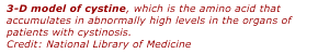 |
Another lysosomal disorder that Dr. Gahl studies is called cystinosis. One of the things lysosomes do in addition to fighting infection is to break down unneeded proteins so their constituent amino acids can be salvaged for future use. In cystinosis, the transporter protein that carries one of these salvaged amino acids, called cystine, out of the lysosome and into the cell cytoplasm is either defective or missing. This causes cystine to build up in the lysosome and form crystals, which end up destroying both the lysosome and the cell around it.
One structure that is ruined early in cystinosis is the proximal tubule of the kidney, which salvages nutrients the body needs from fluid before it is turned into urine and eliminated. The damaged proximal tubules of people with cystinosis cannot do this, so small molecules, such as glucose, amino acids and phosphates spill out of the body and are lost.
Children with the disorder do not grow and have weak, painful bones. If they don't receive treatment, they go into kidney failure and die before age 10. A kidney transplant can prolong life, but the disease can continue to damage other organs, including the eyes, muscles, pancreas and brain.
Dr. Gahl was instrumental in establishing a drug called cysteamine as the treatment of choice for cystinosis. Cysteamine breaks down cystine and keeps it from building up in the tissues, thereby delaying or averting kidney failure, improving growth of children with cystinosis and possibly helping to prevent late complications of the disease. Since coming to the NIH in 1981, Dr. Gahl and his colleagues have seen more than 200 people with cystinosis, and some of his patients who are taking cysteamine are now 40 to 50 years old.
"Cysteamine treatment has changed the course of the disease so dramatically that cystinosis no longer demands the bulk of our efforts," said Dr. Gahl. "It's a very good feeling."
Studying cystinosis has helped the scientific community understand more about kidney function. Other diseases that Dr. Gahl's group explores include: autosomal recessive polycystic kidney disease, which is teaching researchers how kidney and liver cysts form; alkaptonuria, which provides insights about how connective tissue can be destroyed by amino acid breakdowns; hereditary inclusion body myopathy, an enzyme defect that interferes with the maintenance of good muscle tissue; and Hermansky-Pudlak syndrome (HPS), another disorder of the organelles inside cells.
There are eight different types of HPS, each caused by a different genetic defect. People with HPS have albinism because they lack organelles called melanosomes that produce pigment in the hair, eyes and skin. Since melanosome-containing cells nourish the rods and cones in the eyes, people with HPS also have poor vision. A lack of dense bodies in their platelets interferes with blood clotting so they develop bleeding problems, and they have episodes of inflammation in their lungs that cause a build-up of scar tissue called pulmonary fibrosis. For two of the eight types of HPS, the lung disease is fatal. HPS therapy focuses on controlling the bleeding problems and lung damage, and the solutions that are found might be able to help people whose lungs have been scarred by other diseases.
Dr. Gahl emphasizes that researchers, patients and society as a whole are partners in the ongoing challenge of developing new and improved therapies for rare disorders, noting that the impact of such diseases should not be measured by numbers alone. "The test of a society is how it treats its most helpless and disadvantaged members," he said. "People with rare genetic diseases give humanity so much, scientifically and spiritually, that we owe them a huge debt of gratitude. In fact, they make us more human."
Last Reviewed: March 13, 2012


