DNA Microarray Technology
- What is DNA microarray technology?
- What is DNA microarray technology used for?
- How does DNA microarray technology work?
What is DNA microarray technology?
Although all of the cells in the human body contain identical genetic material, the same genes are not active in every cell. Studying which genes are active and which are inactive in different cell types helps scientists to understand both how these cells function normally and how they are affected when various genes do not perform properly. In the past, scientists have only been able to conduct these genetic analyses on a few genes at once. With the development of DNA microarray technology, however, scientists can now examine how active thousands of genes are at any given time.
What is DNA microarray technology used for?
Microarray technology will help researchers to learn more about many different diseases, including heart disease, mental illness and infectious diseases, to name only a few. One intense area of microarray research at the National Institutes of Health (NIH) is the study of cancer. In the past, scientists have classified different types of cancers based on the organs in which the tumors develop. With the help of microarray technology, however, they will be able to further classify these types of cancers based on the patterns of gene activity in the tumor cells. Researchers will then be able to design treatment strategies targeted directly to each specific type of cancer. Additionally, by examining the differences in gene activity between untreated and treated tumor cells - for example those that are radiated or oxygen-starved - scientists will understand exactly how different therapies affect tumors and be able to develop more effective treatments.
How does DNA microarray technology work?

DNA microarrays are created by robotic machines that arrange minuscule amounts of hundreds or thousands of gene sequences on a single microscope slide. Researchers have a database of over 40,000 gene sequences that they can use for this purpose. When a gene is activated, cellular machinery begins to copy certain segments of that gene. The resulting product is known as messenger RNA (mRNA), which is the body's template for creating proteins. The mRNA produced by the cell is complementary, and therefore will bind to the original portion of the DNA strand from which it was copied.
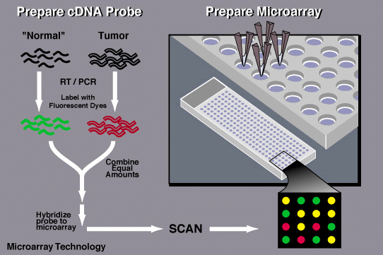
To determine which genes are turned on and which are turned off in a given cell, a researcher must first collect the messenger RNA molecules present in that cell. The researcher then labels each mRNA molecule by using a reverse transcriptase enzyme (RT) that generates a complementary cDNA to the mRNA. During that process fluorescent nucleotides are attached to the cDNA. The tumor and the normal samples are labeled with different fluorescent dyes. Next, the researcher places the labeled cDNAs onto a DNA microarray slide. The labeled cDNAs that represent mRNAs in the cell will then hybridize – or bind – to their synthetic complementary DNAs attached on the microarray slide, leaving its fluorescent tag. A researcher must then use a special scanner to measure the fluorescent intensity for each spot/areas on the microarray slide.
If a particular gene is very active, it produces many molecules of messenger RNA, thus, more labeled cDNAs, which hybridize to the DNA on the microarray slide and generate a very bright fluorescent area. Genes that are somewhat less active produce fewer mRNAs, thus, less labeled cDNAs, which results in dimmer fluorescent spots. If there is no fluorescence, none of the messenger molecules have hybridized to the DNA, indicating that the gene is inactive. Researchers frequently use this technique to examine the activity of various genes at different times. When co-hybridizing Tumor samples (Red Dye) and Normal sample (Green dye) together, they will compete for the synthetic complementary DNAs on the microarray slide. As a result, if the spot is red, this means that that specific gene is more expressed in tumor than in normal (up-regulated in cancer). If a spot is Green, that means that that gene is more expressed in the Normal tissue (Down regulated in cancer). If a spot is yellow that means that that specific gene is equally expressed in normal and tumor.
Last Reviewed: November 15, 2011


