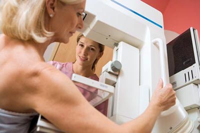
You should have regular clinical breast exams and mammograms to find breast cancer early, when treatment is more likely to work well.

Screening
Mammography
In November 2009, the United States Preventive Services Task Force updated recommendations on breast cancer screening, suggesting that women ages 50 to 74 who are at average risk for getting the disease undergo a routine screening mammogram every two years.
The new recommendations do not advise routine mammography for average-risk women ages 40 to 49.
Self-Examination
The updated 2009 recommendations also advise against teaching breast self-exam (BSE) because no clinical trials to date have shown that teaching of the technique reduces the number of deaths from breast cancer.
According to Dr. Stephen Taplin, senior scientist in NCI's Division of Cancer Control and Population Sciences' Applied Research Program (ARP), this recommendation "certainly does not mean that women shouldn't respond to lumps and bumps or other troublesome changes in their breasts that they discover on their own. Women should go to their healthcare provider when they have a concern."
Clinical Breast Exam
During a clinical breast exam, your healthcare provider inspects your breasts, underarms, and collarbone area. She
- looks for differences in size or shape between the breasts
- checks your skin for a rash, dimpling, or other abnormal signs
- may squeeze your nipples to check for fluid
- uses the pads of her fingers to feel for lumps, pea-sized or larger
- checks the lymph nodes near the breast to see if they are enlarged
If there is a lump, your healthcare provider will feel its size, shape, and texture. She will also see if it moves easily. Lumps that are soft, smooth, round, and movable are likely to be benign. Hard, oddly shaped ones that feel firmly attached within the breast are more likely to be cancer, but you will need further tests to diagnose the problem.
Mammogram
Mammograms are x-ray pictures of breast tissue. They can often show a lump before it can be felt. They also can reveal clusters of tiny specks of calcium. Lumps or specks can be from cancer, precancerous cells, or other conditions. If you have a lump or calcium deposits, you may need further tests to detect the presence of abnormal cells. You should get regular screening mammograms to detect breast cancer early (see Screening for Breast Cancer, next page).
Other Imaging Tests
Ultrasound devices use inaudible sound waves to create images that show whether a breast lump is solid, filled with fluid (a cyst), or a mixture of both. Cysts usually are not cancer. Solid lumps may be. Magnetic resonance imaging (MRI) devices detail the difference between normal and diseased breast tissue.
Biopsy
Biopsies remove small amounts of breast tissue for inspection. They are the only sure way to tell if you have cancer. A pathologist analyzes the tissue or fluid to determine the type of cancer.
Testing Breast Tissue
Special tests on the diseased tissue may help determine treatment:
Hormone receptor tests: Some breast tumors need the hormones estrogen, progesterone, or both, to grow. If they are found, your healthcare provider may recommend hormone therapy.
HER2/neu test: HER2/neu is a protein found on some types of cancer cells. This test shows whether the tissue either has too much HER2/neu protein or too many copies of its gene. If the breast tumor has too much HER2/neu, then targeted therapy, which uses drugs to block the growth of breast cancer cells, may be an option.
