Home » Photos, Images, and Videos » Search Results
Photos, Images, and Videos
Search Results
Instructions for Downloading Photographs and Images
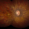 |
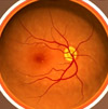
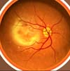
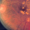
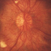
Description: Retina photo of a patient with Leber congenital amaurosis (LCA), an inherited retinal disease that causes severe visual impairment early in childhood. Special gene testing is necessary to determine if the patient has the RPE65-associated type of the disease. Credit: National Eye Institute, National Institutes of Health Ref#: EDA26 |
72
dpi (29M, TIFF) 150 dpi (1M, TIFF) 300 dpi (200K, TIFF) |
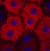 |
Description: Retinal pigment epithelial (RPE) cells stained red by RPE65 antibody. Mutations in the RPE65 gene can cause a form of Leber congenital amaurosis (LCA), an inherited retinal disease that causes severe visual impairment early in childhood. Credit: National Eye Institute, National Institutes of Health Ref#: EDA25 |
72
dpi (2M, TIFF) 150 dpi (7M, TIFF) |
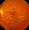 |
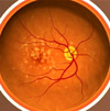
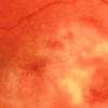
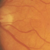
Description: A fundus photo showing intermediate age-related macular degeneration. Credit: National Eye Institute, National Institutes of Health Ref#: EDA22 |
72
dpi (7.5M, TIFF) 150 dpi (14M, TIFF) 300 dpi (14M, TIFF) |
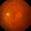 |
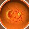
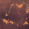
Description: A fundus photo showing geographic atrophy associated with dry age-related macular degeneration. Credit: National Eye Institute, National Institutes of Health Ref#: EDA23 |
72
dpi (3.1M, TIFF) 150 dpi (5M, TIFF) 300 dpi (5M, TIFF) |
 |
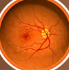
Description: A fundus photo showing neovascular age-related macular degeneration. Credit: National Eye Institute, National Institutes of Health Ref#: EDA24 |
72
dpi (2M, TIFF) 150 dpi (4M, TIFF) 300 dpi (4M, TIFF) |
 |
Description: Neovascular age-related macular degeneration.
Credit: National Eye Institute, National Institutes of Health Ref#: EDA18 |
72
dpi (1.1M, TIFF) |
 |
Description: Intermediate age-related macular degeneration. Credit: National Eye Institute, National Institutes of Health Ref#: EDA19 |
72
dpi (1.1M, TIFF) |
 |
Description: Early intermediate age-related macular degeneration. Credit: National Eye Institute, National Institutes of Health Ref#: EDA20 |
72
dpi (1.1M, TIFF) |
 |
Description: Early age-related macular degeneration. Credit: National Eye Institute, National Institutes of Health Ref#: EDA21 |
72
dpi (1.1M, TIFF) |
 |
Description: Advanced age-related macular degeneration with fibrosis. Credit: National Eye Institute, National Institutes of Health Ref#: EDA17 |
72
dpi (1.1M, TIFF) |
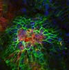 |
Description: Confocal image of rat RPE layer two weeks after laser. A well-defined neovascular membrane labeled with Isolectin Ib4 (red) is detected below proliferating RPE cells (Phalloidin in green). New vessels are growing toward the subretinal space. Credit: National Eye Institute, National Institutes of Health Ref#: EDA15 |
72
dpi (1.8M, TIFF) 150 dpi (1.8M, TIFF) 300 dpi (1.8M, TIFF) |
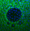 |
Description: Confocal image of rat RPE layer immediately after laser-induced rupture of Bruch’s membrane. A circular disruption of the normal morphology of the choroid, Bruch’s membrane and RPE (green) is visible. DAPI labeled nuclei are blue. Credit: National Eye Institute, National Institutes of Health Ref#: EDA16 |
72
dpi (558K, TIFF) 150 dpi (2.3M, TIFF) 300 dpi (9M, TIFF) |
 |
Description: An acute sudden onset cortical cataract in a person with Type 1 (juvenile) diabetes. Credit: National Eye Institute, National Institutes of Health Ref#: EDA12 |
72
dpi (704K, TIFF) 150 dpi (2.8M, TIFF) |
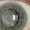 |
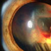
Description: A hypermature age-related cortico-nuclear cataract with a brunescent (brown) nucleus. Credit: National Eye Institute, National Institutes of Health Ref#: EDA13 |
72
dpi (721K, TIFF) 150 dpi (2.9M, TIFF) |
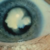 |
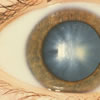
Description: A white congenital cataract. Credit: National Eye Institute, National Institutes of Health Ref#: EDA14 |
72
dpi (354K, TIFF) 150 dpi (1.4M, TIFF) |
 |
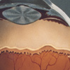
Description: Conventional surgery to treat glaucoma makes a new opening in the meshwork. This new opening helps fluid to leave the eye and lowers intraocular pressure. Pre-surgery image. Credit: National Eye Institute, National Institutes of Health Ref#: EDA11 |
72
dpi (524K, TIFF) 150 dpi (2.1M, TIFF) 300 dpi (8.6M, TIFF) |
 |
Description: Fundus photo showing scatter laser surgery for diabetic retinopathy. Credit: National Eye Institute, National Institutes of Health Ref#: EDA09 |
72
dpi (422K, TIFF) 150 dpi (1.7M, TIFF) 300 dpi (7M, TIFF) |
 |
Description: Fundus photo showing focal laser surgery for diabetic retinopathy. Credit: National Eye Institute, National Institutes of Health Ref#: EDA10 |
72
dpi (424K, TIFF) 150 dpi (1.8M, TIFF) 300 dpi (7.1M, TIFF) |
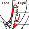 |
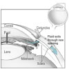
Description: In many people, increased pressure inside the eye causes glaucoma. In the front of the eye is a space called the anterior chamber. A clear liquid flows continuously in and out of this space and nourishes nearby tissues. Credit: National Eye Institute, National Institutes of Health Ref#: EDA02 |
72
dpi (566K, TIFF) 150 dpi (2.4M, TIFF) 300 dpi (9.7M, TIFF) |
 |
Description: Fundus photograph-CMV retinitis Credit: National Eye Institute, National Institutes of Health Ref#: EDA07 |
72
dpi (371K, TIFF) 150 dpi (1.6M, TIFF) 300 dpi (6.3M, TIFF) |
 |
Description: Slit lamp photograph showing retinal detachment in Von Hippel-Lindau disease. Credit: National Eye Institute, National Institutes of Health Ref#: EDA08 |
72
dpi (584K, TIFF) 150 dpi (5M, TIFF) 300 dpi (15M, TIFF) |
 |
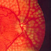
Description: Proliferative retinopathy, an advanced form of diabetic retinopathy, occurs when abnormal new blood vessels and scar tissue form on the surface of the retina. Credit: National Eye Institute, National Institutes of Health Ref#: EDA01 |
72
dpi (370K, TIFF) 150 dpi (1.6M, TIFF) 300 dpi (6.3M, TIFF) |
 |
Description: In background retinopathy, a slight deterioration in the small blood vessels of the retina, portions of the vessels may swell and leak fluid into the surrounding retinal tissue. Credit: National Eye Institute, National Institutes of Health Ref#: EDA03 |
72
dpi (369K, TIFF) 150 dpi (1.6M, TIFF) 300 dpi (6.3M, TIFF) |
 |
Description: Diabetic macular edema. Credit: National Eye Institute, National Institutes of Health Ref#: EDA04 |
72
dpi (369K, TIFF) 150 dpi (1.6M, TIFF) 300 dpi (6.3M, TIFF) |
 |
Description: An artist's conception of the interior of an infant's eye shows the formation of an ROP ridge. Credit: Oregon Health Sciences University Ref#: EDA05 |
72
dpi (372K, TIFF) 150 dpi (1.6M, TIFF) 300 dpi (6.3M, TIFF) |
TOP

