
General Information About Bladder Cancer
Key Points for This Section
- Bladder cancer is a disease in which malignant (cancer) cells form in the tissues of the bladder.
- Smoking can affect the risk of bladder cancer.
- Possible signs of bladder cancer include blood in the urine or pain during urination.
- Tests that examine the urine, vagina, or rectum are used to help detect (find) and diagnose bladder cancer.
- Certain factors affect prognosis (chance of recovery) and treatment options.
Bladder cancer is a disease in which malignant (cancer) cells form in the tissues of the bladder.
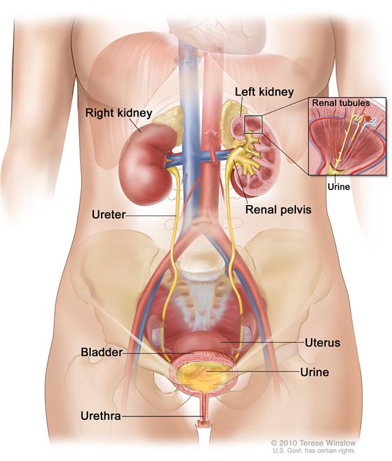
The bladder is a hollow organ in the lower part of the abdomen. It is shaped like a small balloon and has a muscular wall that allows it to get larger or smaller. The bladder stores urine until it is passed out of the body. Urine is the liquid waste that is made by the kidneys when they clean the blood. The urine passes from the two kidneys into the bladder through two tubes called ureters. When the bladder is emptied during urination, the urine goes from the bladder to the outside of the body through another tube called the urethra.

There are three types of bladder cancer that begin in cells in the lining of the bladder. These cancers are named for the type of cells that become malignant (cancerous):
- Transitional cell carcinoma: Cancer that begins in cells in the innermost tissue layer of the bladder. These cells are able to stretch when the bladder is full and shrink when it is emptied. Most bladder cancers begin in the transitional cells.
- Squamous cell carcinoma: Cancer that begins in squamous cells, which are thin, flat cells that may form in the bladder after long-term infection or irritation.
- Adenocarcinoma: Cancer that begins in glandular (secretory) cells that may form in the bladder after long-term irritation and inflammation.
Cancer that is confined to the lining of the bladder is called superficial bladder cancer. Cancer that begins in the transitional cells may spread through the lining of the bladder and invade the muscle wall of the bladder or spread to nearby organs and lymph nodes; this is called invasive bladder cancer.
See the following PDQ summaries for more information:
Smoking can affect the risk of bladder cancer.
Anything that increases your chance of getting a disease is called a risk factor. Having a risk factor does not mean that you will get cancer; not having risk factors doesn't mean that you will not get cancer. Talk to your doctor if you think you may be at risk for bladder cancer. Risk factors for bladder cancer include:
- Using tobacco, especially smoking cigarettes.
- Being exposed to certain substances, such as soot from coal, or chemicals used to make rubber, certain dyes, or textiles.
- Working as a dry cleaner or in places where paper, rope, twine, or clothing is made.
- Taking Aristolochia fangchi, a Chinese herb.
- Drinking water that has high levels of arsenic.
- Having a history of bladder infections, including bladder infections caused by Schistosoma haematobium.
- Using urinary catheters for a long time.
- Having a history of kidney or bladder stones.
- Past treatment with certain anticancer drugs or radiation therapy to the pelvis.
- Having a kidney transplant.
- Having hereditary nonpolyposis colon cancer (HNPCC; Lynch syndrome).
Possible signs of bladder cancer include blood in the urine or pain during urination.
These and other symptoms may be caused by bladder cancer. Other conditions may cause the same symptoms. Check with your doctor if you have any of the following problems:
- Blood in the urine (slightly rusty to bright red in color).
- Frequent urination, or feeling the need to urinate without being able to do so.
- Pain during urination.
- Lower back pain.
Tests that examine the urine, vagina, or rectum are used to help detect (find) and diagnose bladder cancer.
The following tests and procedures may be used:
- Physical exam and history: An exam of the body to check general signs of health, including checking for signs of disease, such as lumps or anything else that seems unusual. A history of the patient’s health habits and past illnesses and treatments will also be taken.
- Internal exam: An exam of the vagina and/or rectum. The doctor inserts gloved fingers into the vagina and/or rectum to feel for lumps.
- Urinalysis: A test to check the color of urine and its contents, such as sugar, protein, red blood cells, and white blood cells.
- Urine cytology: Examination of urine under a microscope to check for abnormal cells.
- Cystoscopy: A procedure to look inside the bladder and urethra to check for abnormal areas. A cystoscope is inserted through the urethra into the bladder. A cystoscope is a thin, tube-like instrument with a light and a lens for viewing. It may also have a tool to remove tissue samples, which are checked under a microscope for signs of cancer.Enlarge
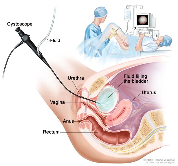 Cystoscopy. A cystoscope (a thin, tube-like instrument with a light and a lens for viewing) is inserted through the urethra into the bladder. Fluid is used to fill the bladder. The doctor looks at an image of the inner wall of the bladder on a computer monitor.
 - Intravenous pyelogram (IVP): A series of x-rays of the kidneys, ureters, and bladder to find out if cancer is present in these organs. A contrast dye is injected into a vein. As the contrast dye moves through the kidneys, ureters, and bladder, x-rays are taken to see if there are any blockages.
- CT scan (CAT scan): A procedure that makes a series of detailed pictures of areas inside the body, taken from different angles. The pictures are made by a computer linked to an x-ray machine. A dye may be injected into a vein or swallowed to help the organs or tissues show up more clearly. This procedure is also called computed tomography, computerized tomography, or computerized axial tomography.
- Biopsy: The removal of cells or tissues so they can be viewed under a microscope by a pathologist to check for signs of cancer. A biopsy for bladder cancer is usually done during cystoscopy. It may be possible to remove the entire tumor during biopsy.
Certain factors affect prognosis (chance of recovery) and treatment options.
The prognosis (chance of recovery) depends on the following:
- The stage of the cancer (whether it is superficial or invasive bladder cancer, and whether it has spread to other places in the body). Bladder cancer in the early stages can often be cured.
- The type of bladder cancer cells and how they look under a microscope.
- The patient’s age and general health.
Treatment options depend on the stage of bladder cancer.
 Back to Top