
General Information about Breast Cancer and Pregnancy
Key Points for This Section
- Breast cancer is a disease in which malignant (cancer) cells form in the tissues of the breast.
- Breast cancer is sometimes detected (found) in women who are pregnant or have just given birth.
- Possible signs of breast cancer include a lump or change in the breast.
- It may be difficult to detect (find) breast cancer early in pregnant or nursing women, whose breasts are often tender and swollen.
- Breast examination should be part of prenatal and postnatal care.
- Tests that examine the breasts are used to detect (find) and diagnose breast cancer.
- If cancer is found, tests are done to study the cancer cells.
- Certain factors affect prognosis (chance of recovery) and treatment options.
Breast cancer is a disease in which malignant (cancer) cells form in the tissues of the breast.
The breast is made up of lobes and ducts. Each breast has 15 to 20 sections called lobes, which have many smaller sections called lobules. The lobes and lobules are connected by thin tubes called ducts.
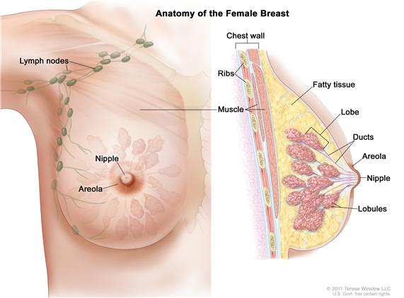
Each breast also contains blood vessels and lymph vessels. The lymph vessels carry an almost colorless fluid called lymph. The lymph vessels lead to small, bean-shaped organs called lymph nodes that help the body fight infection and disease. Lymph nodes are found throughout the body. Clusters of lymph nodes are found near the breast in the axilla (under the arm), above the collarbone, and in the chest.
Breast cancer is sometimes detected (found) in women who are pregnant or have just given birth.
In women who are pregnant or who have just given birth, breast cancer occurs most often between the ages of 32 and 38. Breast cancer occurs about once in every 3,000 pregnancies.
Possible signs of breast cancer include a lump or change in the breast.
Breast cancer may cause any of the following signs and symptoms. Check with your doctor if you have any of the following problems:
- A lump or thickening in or near the breast or in the underarm area.
- A change in the size or shape of the breast.
- A dimple or puckering in the skin of the breast.
- A nipple turned inward into the breast.
- Fluid, other than breast milk, from the nipple, especially if it's bloody.
- Scaly, red, or swollen skin on the breast, nipple, or areola (the dark area of skin that is around the nipple).
- Dimples in the breast that look like the skin of an orange, called peau d’orange.
Other conditions that are not breast cancer may cause these same symptoms.
It may be difficult to detect (find) breast cancer early in pregnant or nursing women, whose breasts are often tender and swollen.
Women who are pregnant, nursing, or have just given birth usually have tender, swollen breasts. This can make small lumps difficult to detect and may lead to delays in diagnosing breast cancer. Because of these delays, cancers are often found at a later stage in these women.
Breast examination should be part of prenatal and postnatal care.
To detect breast cancer, pregnant and nursing women should examine their breasts themselves. Women should also receive clinical breast examinations during their routine prenatal and postnatal examinations.
Tests that examine the breasts are used to detect (find) and diagnose breast cancer.
A doctor should be seen if changes in the breast are noticed. The following tests and procedures may be used:
- Physical exam and history: An exam of the body to check general signs of health, including checking for signs of disease, such as lumps or anything else that seems unusual. A history of the patient’s health habits and past illnesses and treatments will also be taken.
- Clinical breast exam (CBE): An exam of the breast by a doctor or other health professional. The doctor will carefully feel the breasts and under the arms for lumps or anything else that seems unusual.
- MRI (magnetic resonance imaging): A procedure that uses a magnet, radio waves, and a computer to make a series of detailed pictures of areas inside the body. This procedure is also called nuclear magnetic resonance imaging (NMRI).
- Ultrasound exam: A procedure in which high-energy sound waves (ultrasound) are bounced off internal tissues or organs and make echoes. The echoes form a picture of body tissues called a sonogram.
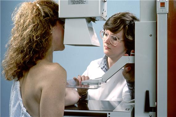
- Mammogram: An x-ray of the breast. A mammogram can be performed with little risk to the fetus. Mammograms in pregnant women may appear negative even though cancer is present.
- Blood chemistry studies: A procedure in which a blood sample is checked to measure the amounts of certain substances released into the blood by organs and tissues in the body. An unusual (higher or lower than normal) amount of a substance can be a sign of disease in the organ or tissue that makes it.
- Biopsy: The removal of cells or tissues so they can be viewed under a microscope by a pathologist to check for signs of cancer. If a lump in the breast is found, the doctor may need to remove a small piece of the lump. Four types of biopsies are as follows:
- Excisional biopsy: The removal of an entire lump of tissue.
- Incisional biopsy: The removal of part of a lump or a sample of tissue.
- Core biopsy: The removal of tissue using a wide needle.
- Fine-needle aspiration (FNA) biopsy: The removal of tissue or fluid, using a thin needle.
If cancer is found, tests are done to study the cancer cells.
Decisions about the best treatment are based on the results of these tests. The tests give information about:
- How quickly the cancer may grow.
- How likely it is that the cancer will spread through the body.
- How well certain treatments might work.
- How likely the cancer is to recur (come back).
Tests include the following:
- Estrogen and progesterone receptor test: A test to measure the amount of estrogen and progesterone (hormones) receptors in cancer tissue. If there are more estrogen and progesterone receptors than normal, the cancer may grow more quickly. The test results show whether treatment to block estrogen and progesterone may stop the cancer from growing.
- Human epidermal growth factor type 2 receptor (HER2/neu) test: A laboratory test to measure how many HER2/neu genes there are and how much HER2/neu protein is made in a sample of tissue. If there are more HER2/neu genes or higher levels of HER2/neu protein than normal, the cancer may grow more quickly and is more likely to spread to other parts of the body. The cancer may be treated with drugs that target the HER2/neu protein, such as trastuzumab (Herceptin) and lapatinib (Tykerb).
- Multigene tests: Tests in which samples of tissue are studied to look at the activity of many genes at the same time. These tests may help predict whether cancer will spread to other parts of the body or recur (come back).
- Oncotype DX: This test helps predict whether stage I or stage II breast cancer that is estrogen receptor positive and node-negative will spread to other parts of the body. If the risk of the cancer spreading is high, chemotherapy may be given to lower the risk.
- MammaPrint: This test helps predict whether stage I or stage II breast cancer that is node-negative will spread to other parts of the body. If the risk of the cancer spreading is high, chemotherapy may be given to lower the risk.
Certain factors affect prognosis (chance of recovery) and treatment options.
The prognosis (chance of recovery) and treatment options depend on the following:
- The stage of the cancer (whether it is in the breast only or has spread to other places in the body).
- The size of the tumor.
- The type of breast cancer.
- The age of the fetus.
- Whether there are symptoms.
- The patient’s general health.
 Back to Top