
General Information About Pancreatic Neuroendocrine Tumors (Islet Cell Tumors)
Key Points for This Section
- Pancreatic neuroendocrine tumors form in hormone-making cells (islet cells) of the pancreas.
- Pancreatic NETs may or may not cause symptoms.
- There are different kinds of functional pancreatic NETs.
- Having certain syndromes can increase the risk of pancreatic NETs.
- Different types of pancreatic NETs have different signs and symptoms.
- Lab tests and imaging tests are used to detect (find) and diagnose pancreatic NETs.
- Other kinds of lab tests are used to check for the specific type of pancreatic NETs.
- Certain factors affect prognosis (chance of recovery) and treatment options.
Pancreatic neuroendocrine tumors form in hormone-making cells (islet cells) of the pancreas.
The pancreas is a gland about 6 inches long that is shaped like a thin pear lying on its side. The wider end of the pancreas is called the head, the middle section is called the body, and the narrow end is called the tail. The pancreas lies behind the stomach and in front of the spine.
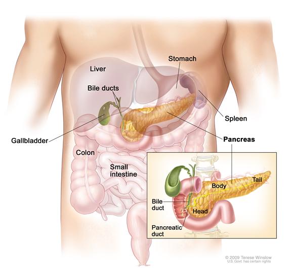
There are two kinds of cells in the pancreas:
- Endocrine pancreas cells make several kinds of hormones (chemicals that control the actions of certain cells or organs in the body), such as insulin to control blood sugar. They cluster together in many small groups (islets) throughout the pancreas. Endocrine pancreas cells are also called islet cells or islets of Langerhans.
- Exocrine pancreas cells make enzymes that are released into the small intestine to help the body digest food. Most of the pancreas is made of ducts with small sacs at the end of the ducts, which are lined with exocrine cells. This summary discusses islet cell tumors of the endocrine pancreas. See the PDQ summary on Pancreatic Cancer Treatment for information on exocrine pancreatic cancer.
A pancreatic neuroendocrine tumor (NET) may also be called a pancreatic endocrine tumor (PET), islet cell tumor, islet cell carcinoma, or pancreatic carcinoid.
Pancreatic NETs are much less common than pancreatic exocrine tumors and have a better prognosis.
Pancreatic NETs may or may not cause symptoms.
Pancreatic NETs may be functional (the hormones that are released cause symptoms) or nonfunctional (the hormones that are released do not cause symptoms) tumors:
- Functional tumors make one or more hormones, such as gastrin, insulin, and glucagon, that cause symptoms. Most functional tumors are benign (not cancer).
- Nonfunctional tumors make substances that do not cause symptoms. Symptoms are caused by the tumor as it spreads and grows. Most nonfunctional tumors are malignant (cancer).
Most pancreatic NETs are functional tumors.
There are different kinds of functional pancreatic NETs.
Pancreatic NETs make different kinds of hormones such as gastrin, insulin, and glucagon. Functional pancreatic NETs include the following:
- Gastrinoma: A tumor that forms in cells that make gastrin. Gastrin is a hormone that causes the stomach to release an acid that helps digest food. Both gastrin and stomach acid are increased by gastrinomas. When increased stomach acid, stomach ulcers, and diarrhea are caused by a tumor that makes gastrin, it is called Zollinger-Ellison syndrome. A gastrinoma usually forms in the head of the pancreas and sometimes forms in the small intestine. Most gastrinomas are malignant (cancer).
- Insulinoma: A tumor that forms in cells that make insulin. Insulin is a hormone that controls the amount of glucose (sugar) in the blood. It moves glucose into the cells, where it can be used by the body for energy. Insulinomas are usually slow-growing tumors that rarely spread. An insulinoma forms in the head, body, or tail of the pancreas. Insulinomas are usually benign (not cancer).
- Glucagonoma: A tumor that forms in cells that make glucagon. Glucagon is a hormone that increases the amount of glucose in the blood. It causes the liver to break down glycogen. Too much glucagon causes hyperglycemia (high blood sugar). A glucagonoma usually forms in the tail of the pancreas. Most glucagonomas are malignant (cancer).
- Other types of tumors: There are other rare types of functional pancreatic NETs that make hormones, including hormones that control the balance of sugar, salt, and water in the body. These tumors include:
- VIPomas, which make vasoactive intestinal peptide. VIPoma may also be called Verner-Morrison syndrome.
- Somatostatinomas, which make somatostatin.
Having certain syndromes can increase the risk of pancreatic NETs.
Anything that increases your risk of getting a disease is called a risk factor. Having a risk factor does not mean that you will get cancer; not having risk factors doesn't mean that you will not get cancer. People who think they may be at risk should discuss this with their doctor.
Multiple endocrine neoplasia type 1 (MEN1) syndrome is a risk factor for pancreatic NETs.
Different types of pancreatic NETs have different signs and symptoms.
Symptoms can be caused by the growth of the tumor and/or by hormones the tumor makes. Some tumors may not cause symptoms. Conditions other than pancreatic NETs can cause the symptoms listed below. Talk to your doctor if any of these problems occur.
Signs and symptoms of a non-functional pancreatic NET
A non-functional pancreatic NET may grow for a long time without causing symptoms. It may grow large or spread to other parts of the body before it causes symptoms, such as:
- Diarrhea.
- Indigestion.
- A lump in the abdomen.
- Pain in the abdomen or back.
- Yellowing of the skin and whites of the eyes.
Signs and symptoms of a functional pancreatic NET
The symptoms of a functional pancreatic NET depend on the type of hormone being made.
Too much gastrin may cause:
- Stomach ulcers that keep coming back.
- Pain in the abdomen, which may spread to the back. The pain may come and go and it may go away after taking an antacid.
- The flow of stomach contents back into the esophagus (gastroesophageal reflux).
- Diarrhea.
Too much insulin may cause:
- Low blood sugar. This can cause blurred vision, headache, and feeling lightheaded, tired, weak, shaky, nervous, irritable, sweaty, confused, or hungry.
- Fast heartbeat.
Too much glucagon may cause:
- Skin rash on the face, stomach, or legs.
- High blood sugar. This can cause headaches, frequent urination, dry skin and mouth, or feeling hungry, thirsty, tired, or weak.
- Blood clots. Blood clots in the lung can cause shortness of breath, cough, or pain in the chest. Blood clots in the arm or leg can cause pain, swelling, warmth, or redness of the arm or leg.
- Diarrhea.
- Weight loss for no known reason.
- Sore tongue or sores at the corners of the mouth.
Too much vasoactive intestinal peptide (VIP) may cause:
- Very large amounts of watery diarrhea.
- Dehydration. This can cause feeling thirsty, making less urine, dry skin and mouth, headaches, dizziness, or feeling tired.
- Low potassium level in the blood. This can cause muscle weakness, aching, or cramps, numbness and tingling, frequent urination, fast heartbeat, and feeling confused or thirsty.
- Cramps or pain in the abdomen.
- Weight loss for no known reason.
Too much somatostatin may cause:
- High blood sugar. This can cause headaches, frequent urination, dry skin and mouth, or feeling hungry, thirsty, tired, or weak.
- Diarrhea.
- Steatorrhea (very foul-smelling stool that floats).
- Gallstones.
- Yellowing of the skin and whites of the eyes.
- Weight loss for no known reason.
Lab tests and imaging tests are used to detect (find) and diagnose pancreatic NETs.
The following tests and procedures may be used:
- Physical exam and history: An exam of the body to check general signs of health, including checking for signs of disease, such as lumps or anything else that seems unusual. A history of the patient’s health habits and past illnesses and treatments will also be taken.
- Blood chemistry studies: A procedure in which a blood sample is checked to measure the amounts of certain substances, such as glucose (sugar), released into the blood by organs and tissues in the body. An unusual (higher or lower than normal) amount of a substance can be a sign of disease in the organ or tissue that makes it.
- Immunohistochemistry study: A laboratory test in which a substance such as an antibody, dye, or radioisotope is added to a sample of cancer tissue to test for certain antigens. This type of study is used to tell the difference between different types of cancer.
- Abdominal CT scan (CAT scan): A procedure that makes a series of detailed pictures of the abdomen, taken from different angles. The pictures are made by a computer linked to an x-ray machine. A dye may be injected into a vein or swallowed to help the organs or tissues show up more clearly. This procedure is also called computed tomography, computerized tomography, or computerized axial tomography.
- MRI (magnetic resonance imaging): A procedure that uses a magnet, radio waves, and a computer to make a series of detailed pictures of areas inside the body. This procedure is also called nuclear magnetic resonance imaging (NMRI).
- Somatostatin receptor scintigraphy: A type of radionuclide scan that may be used to find small pancreatic NETs. A small amount of radioactive octreotide (a hormone that attaches to tumors) is injected into a vein and travels through the blood. The radioactive octreotide attaches to the tumor and a special camera that detects radioactivity is used to show where the tumors are in the body. This procedure is also called octreotide scan and SRS.
- Abdominal ultrasound: An ultrasound exam used to make pictures of the inside of the abdomen. The ultrasound transducer is pressed against the skin of the abdomen and directs high-energy sound waves (ultrasound) into the abdomen. The sound waves bounce off the internal tissues and organs and make echoes. The transducer receives the echoes and sends them to a computer, which uses the echoes to make pictures called sonograms. The picture can be printed to be looked at later.
- Endoscopic ultrasound (EUS): A procedure in which an endoscope is inserted into the body, usually through the mouth or rectum. An endoscope is a thin, tube-like instrument with a light and a lens for viewing. A probe at the end of the endoscope is used to bounce high-energy sound waves (ultrasound) off internal tissues or organs and make echoes. The echoes form a picture of body tissues called a sonogram. This procedure is also called endosonography.
- Angiogram: A procedure to look at blood vessels and the flow of blood. A contrast dye is injected into the blood vessel. As the contrast dye moves through the blood vessel, x-rays are taken to see if there are any blockages.
- Laparotomy: A surgical procedure in which an incision (cut) is made in the wall of the abdomen to check the inside of the abdomen for signs of disease. The size of the incision depends on the reason the laparotomy is being done. Sometimes organs are removed or tissue samples are taken and checked under a microscope for signs of disease.
- Intraoperative ultrasound: A procedure that uses high-energy sound waves (ultrasound) to create images of internal organs or tissues during surgery. A transducer placed directly on the organ or tissue is used to make the sound waves, which create echoes. The transducer receives the echoes and sends them to a computer, which uses the echoes to make pictures called sonograms.
- Biopsy: The removal of cells or tissues so they can be viewed under a microscope by a pathologist to check for signs of cancer. There are several ways to do a biopsy for pancreatic NETs. Cells may be removed using a fine or wide needle inserted into the pancreas during an x-ray or ultrasound. Tissue may also be removed during a laparoscopy (a surgical incision made in the wall of the abdomen).
- Bone scan: A procedure to check if there are rapidly dividing cells, such as cancer cells, in the bone. A very small amount of radioactive material is injected into a vein and travels through the blood. The radioactive material collects in bones where cancer cells have spread and is detected by a scanner.
Other kinds of lab tests are used to check for the specific type of pancreatic NETs.
The following tests and procedures may be used:
Gastrinoma
- Fasting serum gastrin test: A test in which a blood sample is checked to measure the amount of gastrin in the blood. This test is done after the patient has had nothing to eat or drink for at least 8 hours. Conditions other than gastrinoma can cause an increase in the amount of gastrin in the blood.
- Gastric acid secretion test: A test to measure the amount of acid made by the stomach. A tube is inserted through the nose or throat, into the stomach. Gastrin or insulin is injected into the patient, which causes the stomach to make stomach secretions (gastric acid). Four samples of gastric acid are taken through the tube 15 minutes apart. These four samples are used to find out the lowest and highest amounts of gastric acid made during the test and the pH level of the gastric secretions.
- Secretin stimulation test: If the gastric acid secretion test result is not normal, a secretin stimulation test may be done. The tube is moved into the small intestine and samples are taken from the small intestine after a drug called secretin is injected. Secretin causes the small intestine to make acid. When there is a gastrinoma, the secretin causes an increase in how much gastric acid is made and the level of gastrin in the blood.
- Calcium infusion test: A test to measure the amount of gastrin in the blood after a drug called calcium gluconate is infused. Blood samples will be taken to measure the amount of gastrin in the blood at set times.
- Somatostatin receptor scintigraphy: A type of radionuclide scan that may be used to find small pancreatic NETs. A small amount of radioactive octreotide (a hormone that attaches to tumors) is injected into a vein and travels through the blood. The radioactive octreotide attaches to the tumor and a special camera that detects radioactivity is used to show where the tumors are in the body. This procedure is also called octreotide scan and SRS.
Insulinoma
- Fasting serum glucose and insulin test: A test in which a blood sample is checked to measure the amounts of glucose (sugar) and insulin in the blood. The test is done after the patient has had nothing to eat or drink for at least 24 hours.
- C-peptide suppression test: A test in which a blood sample is checked to measure the amount of C-peptide in the blood. Insulin is injected into a vein to lower the patient’s blood sugar. This should decrease the amount of insulin and C-peptide that the body releases into the blood. In patients who have insulinoma, the insulin and C-peptide levels do not drop because the tumor is also releasing insulin and C-peptide into the blood.
Glucagonoma
- Fasting serum glucagon test: A test in which a blood sample is checked to measure the amount of glucagon in the blood. The test is done after the patient has had nothing to eat or drink for at least 8 hours.
Other tumor types
- VIPoma
- Serum VIP (vasoactive intestinal peptide) test: A test in which a blood sample is checked to measure the amount of VIP.
- Blood chemistry studies: A procedure in which a blood sample is checked to measure the amounts of certain substances released into the blood by organs and tissues in the body. An unusual (higher or lower than normal) amount of a substance can be a sign of disease in the organ or tissue that makes it. In VIPoma, there is a lower than normal amount of potassium.
- Stool analysis: A stool sample is checked for a higher than normal sodium (salt) and potassium levels.
- Somatostatinoma
- Fasting serum somatostatin test: A test in which a blood sample is checked to measure the amount of somatostatin in the blood. The test is done after the patient has had nothing to eat or drink for at least 8 hours.
- Somatostatin receptor scintigraphy: A type of radionuclide scan that may be used to find small pancreatic NETs. A small amount of radioactive octreotide (a hormone that attaches to tumors) is injected into a vein and travels through the blood. The radioactive octreotide attaches to the tumor and a special camera that detects radioactivity is used to show where the tumors are in the body. This procedure is also called octreotide scan and SRS.
Certain factors affect prognosis (chance of recovery) and treatment options.
Pancreatic NETs can often be cured. The prognosis (chance of recovery) and treatment options depend on the following:
- The type of cancer cell.
- Where the tumor is found in the pancreas.
- Whether the tumor has spread to more than one place in the pancreas or to other parts of the body.
- Whether the patient has MEN1 syndrome.
- The patient's age and general health.
- Whether the cancer has just been diagnosed or has recurred (come back).
 Back to Top