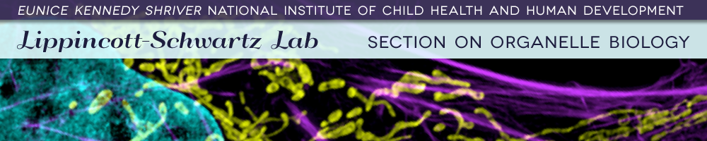


The Lippincott-Schwartz group combines cell biology, biochemical and biophysical approaches to investigate the interplay between membrane organelles, cytoskeleton and metabolism in cell organization and function. To achieve this, the lab comprises an interdisciplinary, international team with expertise in cell biology, physics, chemistry, mathematical modeling, engineering and computer science. Our research relies heavily on microscopy, including cutting edge fluorescence-based technologies to study questions at different spatial scales. We also employ biochemistry, in vitro reconstitution and mathematical modeling to develop and test various mechanistic hypotheses. At the small scale, we employ the super-resolution imaging techniques of photoactivated localization microscopy (PALM), interferometric 3D PALM, single particle tracking PALM and pair correlation PALM to map out the spatial organization, stoichiometry and dynamics of proteins associated with different membrane-bound compartments and the cytoskeleton. We also employ fluorescence photobleaching, photoactivation, fluorescence correlation and fluorescence energy transfer methods to measure protein-protein interactions, protein turnover rates and protein association rates. These approaches allow us to assay cellular functions at the molecular scale in living cells.
At the larger scale, we are investigating how complex behaviors of cells arise, such as cell crawling, polarization, furrowing, cytokinesis, cell fate determination, viral budding and intercellular transfer. These intricate behaviors are studied by quantitatively analyzing diverse intracellular processes, including membrane trafficking, autophagy, actin/microtubule dynamics, and organelle assembly/disassembly pathways, which undergo dramatic changes as cells alter their behavior and organization throughout life. To assist with these efforts, different fluorescence-based imaging approaches, including TIRF imaging, spinning disk and laser scanning confocal microscopy, are combined with FRAP, FLIP and photoactivation to obtain large image data sets. The data sets are computationally processed to extract biochemical and biophysical parameters (which can be related to results from conventional biochemical assays). The results are then used to generate mechanistic understanding and predictive models of the behavior of cells and subcellular structures (including ER, Golgi, cilia, endosomes, lysosomes, autophagosomes and mitochondria) both under healthy and pathological conditions.
Opportunities:
Please contact Jennifer Lippincott-Schwartz at jlippin@helix.nih.gov![]() for information regarding the availability of post-doctoral and graduate fellowship positions in the lab. Graduate students may apply through the Graduate Partnership Program
for information regarding the availability of post-doctoral and graduate fellowship positions in the lab. Graduate students may apply through the Graduate Partnership Program![]() that sponsors doctoral students at NIH through partnerships with various Universities, including University of Maryland, Johns Hopkins University
that sponsors doctoral students at NIH through partnerships with various Universities, including University of Maryland, Johns Hopkins University![]() , Oxford University (UK), or Cambridge University (UK)
, Oxford University (UK), or Cambridge University (UK)![]() .
.
Mailing Address: Cell Biology and Metabolism BranchNational Institutes of Health |
(Click Map To Enlarge)
|