Novel Breast CT Scanner Enhances Ability to Discern Tumors
Technique doesn’t require painful breast compression
 |
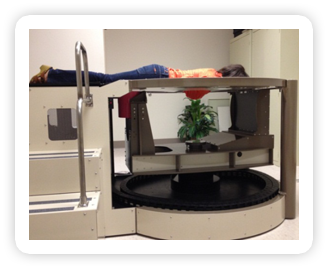
| Figure 1: Boone’s dedicated breast CT scanner |
An NIBIB grantee has developed a dedicated breast CT scanner that allows the breast to be imaged in three dimensions and could help radiologists detect hard-to-find tumors. The scanner uses a radiation dose comparable to standard x-ray mammography and doesn’t require compression of the breast. With support from NIBIB, John Boone, PhD, and his research team at University of California, Davis, are now adding new imaging capabilities to the dedicated breast CT scanner previously developed with NIBIB funding. They have recently added positron emission tomography imaging (PET) and contrast-enhanced CT, which will improve the scanner’s ability to image tumors. In addition, they are collaborating with other researchers to develop computer-aided detection software that will help radiologists interpret the vast amount of data generated by these new techniques. Though currently only used for research purposes, these experimental breast CT scanners will likely be ready for clinical use after clinical trials are concluded and FDA marketing approval is obtained.
Background
For the past 40 years, x-ray mammography has been the standard imaging technique used to detect breast cancer; yet this life-saving technology is not without limitations. Sensitivity for cancer detection among women using this technique is around 80%, meaning a tumor is likely to be missed in about 20% of women who have cancer. Additionally, 10% of women who go in for a routine mammography are called back for additional testing. Of those, 90% walk away with a clean bill of health, but not before enduring significant emotional stress.1
The biggest factor contributing to decreased sensitivity and increased recall rates is the fact that mammography produces a two dimensional image of a three dimensional object. In mammography, low energy x-rays pass through a compressed breast to produce an image; generally both a top and side view are taken. Because x-rays travel easily through areas of low density such as fatty tissue, these areas of the breast appear translucent. Tumors, which have higher densities, appear white against a dark background. However, in addition to fat, the breast is also comprised of glandular and connective tissue, which, because of its higher density, also appears white against a dark background. For women with dense breasts (usually younger women), small tumors can be obscured behind layers of dense tissue. This is less of an issue with older women, since breast density tends to decline as women go through menopause.
Creating a Dedicated Breast CT Scanner
Earlier in his career, Boone recognized the potential for CT to help radiologists detect breast cancer.
“I’m a professor…and when I give lectures on CT, what I basically teach is that CT has better contrast resolution than mammography,” said Boone. “It became a natural conclusion that if you really wanted to improve contrast resolution for detecting breast cancer, CT was the way to go.”
In a CT scanner, an x-ray tube and detector—positioned on opposite sides of a patient--rotate 360 degrees while sending x-rays through the body at many different angles. Each rotation generates a series of cross-sectional images or “slices” and multiple slices are acquired along the length of the subject while moving through the scanner. The slices are then reconstructed by a computer to generate a composite 3D image that radiologists can view as an entire volume or as component slices.
Although CT resolution is quite high, when Boone first announced his intention to build a CT scanner for breast cancer detection, his colleagues were skeptical. The reason is that a conventional CT scan of the chest requires a hefty dose of radiation because x-rays have to penetrate a patient’s entire thorax in order to reach the x-ray detector located on the opposite side. To Boone’s peers, it was inevitable that a CT scan of the breast would do more harm than good.
But Boone, a medical physicist and Vice Chair of Radiology and Professor of Radiology and Biomedical Imaging at UC Davis, was determined. He believed he could decrease a patient’s radiation dose by scanning only a woman’s breast and not her chest.
Boone’s “Dedicated Breast CT” scanner requires a woman to lie prone on a specially designed large table with her breast suspended through a hole in the middle (Figure 1). The scanner rotates only around the breast, taking hundreds of x-rays without passing any through the chest. An added bonus: compression of the breast—often painful for women—isn’t necessary.
In 2001, using a computer simulation, Boone demonstrated that his proposed CT scanner would be able to successfully produce a 3D image of the breast using a radiation dose comparable to a standard mammogram.
This was a turning point for Boone and with the help of funding from NIBIB, he began to construct his breast CT scanner. In November of 2004, Boone and his colleagues became the first lab to use a CT breast scanner on a human subject.
As predicted, results from early clinical trials confirmed that breast CT was superior to traditional mammography for the visualization of tumors. However, mammography was still better at detecting microcalcifications which can signal the presence a specific type of cancer called ductal carcinoma in situ (DCIS).
Future developments
To date, Boone and his lab have developed three breast CT scanner prototypes (they are currently working on a fourth), and his scanners have imaged over 600 women in clinical trials. With support from NIBIB, Boone has continued to optimize his scanners by integrating imaging modalities such as positron emission tomography (PET) and contrast-enhanced CT.
In PET imaging, a patient is given an injection of a radioactive sugar molecule that accumulates in high metabolic areas such as tumors. After 45 minutes of uptake, the patient is scanned and emissions from the sugar molecules can be detected to determine whether a tumor is present. Boone says the combination of PET with high-resolution anatomical images generated by CT is a powerful diagnostic tool.
 |
| Figure 2: Breast image generated with dbPET/CT. Orange and purple represent areas of increased metabolic activity. |
“We colorize the PET scan and lay it onto the gray-scale breast CT image,” said Boone. “It provides a pretty dramatic image of the tumor.” (Figure 2)
Boone hopes that contrast-enhanced CT—a method in which a woman is injected with a non-radioactive dye that lights up areas dense with blood vessels such as tumors-- may improve breast CT’s ability to detect microcalcifications and help distinguish between benign and malignant tumors.
In addition to adding more imaging capabilities, Boone, in collaboration with researchers at the University of Chicago, is also developing software for computer-aided detection. “One of the issues we recognized early on is that we’re essentially asking radiologists to move from looking at two images per breast to looking at about 500 images per breast,” said Boone. “Radiologists are busy and the extra time required would likely preclude the deployment of such a device.” The goal is to produce software that uses algorithms to automatically read those 500 images and classify any detected tumors as benign or malignant, a process that could both save time and improve the accuracy of diagnoses.
Clinical Use
Boone’s breast CT scanners are currently only used for research purposes. However, several companies worldwide are developing breast CT scanners similar to his prototypes for clinical use. Boone says he wouldn’t be surprised to find a CT scanner in the clinic within the decade.
“The companies have to go through the [FDA] approval process, which is quite lengthy, but it would be realistic to think that breast CT could be available in perhaps five to eight years in the United States,” said Boone.
-- Margot Kern
1 Performance measures for 3,603,832 screening mammography studies from 1996 to 2006 by age. Rockville, MD: National Cancer Institute; 2008. NCI-funded breast cancer surveillance consortium co-operative agreement.
 |
| Dr. John Boone |
Last Updated On 01/28/2013