
Copyright © 1993-2013, University of Washington, Seattle. All rights reserved.
NCBI Bookshelf. A service of the National Library of Medicine, National Institutes of Health.
Pagon RA, Bird TD, Dolan CR, et al., editors. GeneReviews™ [Internet]. Seattle (WA): University of Washington, Seattle; 1993-.
Bookshelf ID: NBK114806PMID: 23236640
Summary
Disease characteristics. KAT6B-related disorders include genitopatellar syndrome (GPS) and Say-Barber-Biesecker variant of Ohdo syndrome (Ohdo syndrome, SBBYS variant, or SBBYSS [Say-Barber-Biesecker-Young-Simpson syndrome]). Both phenotypes are characterized by significant global developmental delay/intellectual disability, hypotonia, genital abnormalities in males (cryptorchidism), and patellar hypoplasia/agenesis. Congenital heart defects, dental anomalies, hearing loss, and thyroid anomalies are common to both phenotypes. Also observed in GPS are flexion contractures of the hips and knees, club feet, agenesis of the corpus callosum with microcephaly, and hydronephrosis and/or multiple renal cysts. In SBBYS lower extremity joint stiffness, long thumbs/great toes, immobile mask-like face, blepharophimosis/ptosis, and lacrimal duct anomalies are observed.
Diagnosis/testing. The diagnosis of KAT6B-related disorders is based on clinical findings consistent with the GPS-SBBYSS group of disorders and presence of a heterozygous mutation in KAT6B. Molecular genetic testing is available on a clinical basis.
Management. Treatment of manifestations: Educational intervention and speech therapy beginning in infancy. Orthopedic intervention as needed for contractures and club foot and physical therapy to help increase joint mobility. Routine management of cryptorchidism, congenital heart defects, dental anomalies, hearing loss, thyroid anomalies, and ophthalmologic findings.
Surveillance: Yearly evaluations of developmental progress, contractures and/or scoliosis by an orthopedist, ophthalmologic problems such as amblyopia (in SBBYSS), thyroid function tests, heart defects, and kidneys if hydronephrosis and/or multiple renal cysts are present
Genetic counseling. KAT6B-related disorders are inherited in an autosomal dominant manner. To date, most individuals with a KAT6B-related disorder have had a de novo mutation. Prenatal diagnosis is possible for families in which the disease-causing mutation has been identified.
Diagnosis
KAT6B-related disorders include genitopatellar syndrome (GPS) and Say-Barber-Biesecker variant of Ohdo syndrome (SBBYSS [Say-Barber-Biesecker-Young-Simpson syndrome]), which can also be referred to as the GPS-SBBYSS group of disorders.
Genitopatellar syndrome (GPS). While clinical diagnostic criteria have not been defined for genitopatellar syndrome, the authors propose that the following features (Table 1) should prompt KAT6B molecular genetic testing. Individuals with two major features or one major feature and two minor features are likely to have a KAT6B mutation.
Table 1. Indicative Guide for Testing for GPS
| Category | Features |
|---|---|
| Major features | • Genital anomalies (females: clitoromegaly and/or hypoplasia of the labia minora or majora; males: cryptorchidism and scrotal hypoplasia) • Patellar hypoplasia/agenesis • Flexion contractures at the hips and knees (including club feet) • Agenesis of the corpus callosum with microcephaly • Hydronephrosis and/or multiple renal cysts |
| Minor features | • Congenital heart defect • Dental anomalies (delayed eruption of teeth) • Hearing loss • Thyroid anomalies • Anal anomalies • Hypotonia • Global developmental delay/intellectual disability |
Say-Barber-Biesecker variant of Ohdo syndrome (SBBYSS). Criteria for the clinical diagnosis of SBBYSS proposed by White et al [2003] included the mandatory findings of blepharophimosis, ptosis, and intellectual disability and the supporting findings of depressed nasal bridge, hypoplastic teeth, deafness, undescended testes, and hypotonia. The authors propose slightly broader features (Table 2) to prompt KAT6B molecular genetic testing in individuals suspected of having SBBYSS. Individuals with two major features or one major feature and two minor features are likely to have a KAT6B mutation.
Table 2. Indicative Guide for Testing for Ohdo/SBBYS Syndrome (SBBYSS)
| Category | Features |
|---|---|
| Major features | • Long thumbs/great toes • Immobile mask-like face • Blepharophimosis/ptosis • Lacrimal duct anomalies • Patellar hypoplasia/agenesis |
| Minor features | • Congenital heart defect • Dental anomalies (hypoplastic teeth and/or delayed eruption of teeth) • Hearing loss • Thyroid anomalies • Cleft palate • Genital anomalies (males: cryptorchidism) • Hypotonia • Global developmental delay/intellectual disability |
Testing
Cytogenetic. Karyotyping is clinically available and may be useful; one individual had a “Noonan syndrome-like” phenotype resulting from a translocation interrupting intron 3 of KAT6B [Kraft et al 2011].
Molecular Genetic Testing
Gene. KAT6B-related disorders are caused by mutations in KAT6B.
Table 3. Summary of Molecular Genetic Testing Used in KAT6B-Related Disorders
| Gene Symbol | Test Method | Mutations Detected | Mutation Detection Frequency by Test Method 1 | Test Availability |
|---|---|---|---|---|
| KAT6B | Sequence analysis | Sequence variants 2 | 11/12 3 | Clinical |
| Deletion / duplication analysis 4 | Exonic or whole-gene deletions | Unknown, none reported | Research only 5 |
Test Availability refers to availability in the GeneTests™ Laboratory Directory. GeneReviews designates a molecular genetic test as clinically available only if the test is listed in the GeneTests™ Laboratory Directory by either a US CLIA-licensed laboratory or a non-US clinical laboratory. GeneTests does not verify laboratory-submitted information or warrant any aspect of a laboratory's licensure or performance. Clinicians must communicate directly with the laboratories to verify information.
1. The ability of the test method used to detect a mutation that is present in the indicated gene
2. Examples of mutations detected by sequence analysis may include small intragenic deletions/insertions and missense, nonsense, and splice site mutations; typically, exonic or whole-gene deletions/duplications are not detected.
3. Campeau et al [2012a], Simpson et al [2012]
4. Testing that identifies deletions/duplications not readily detectable by sequence analysis of the coding and flanking intronic regions of genomic DNA; included in the variety of methods that may be used are: quantitative PCR, long-range PCR, multiplex ligation-dependent probe amplification (MLPA), and chromosomal microarray (CMA) that includes this gene/chromosome segment. See CMA.
5. No laboratories offering clinical testing for this gene are listed in the GeneTests™ Laboratory Directory; clinical confirmation of mutations identified in a research laboratory may be available. See
.
Interpretation of test results. For issues to consider in interpretation of sequence analysis results, click here.
Information on specific allelic variants may be available in Molecular Genetics (see Table A. Genes and Databases and/or Pathologic allelic variants).
Testing Strategy
To confirm/establish the diagnosis in a proband
For individuals with features suggestive of GPS:
Sequence analysis is appropriate, with particular focus on KAT6B exon 18 because all known GPS-causing mutations have occurred in the proximal coding region of that exon.
If no mutation is found, sequence analysis of the rest of the gene can be performed; if no mutation is found, cytogenetic analysis can be performed.
The yield for deletion/ duplication analysis is unknown as such mutations have not been reported as a cause of GPS.
For individuals with features suggestive of SBBYSS:
Sequence analysis of the entire gene should be performed (starting with exon 18); if no mutation is found, cytogenetic analysis can be performed.
The yield for deletion/ duplication analysis is unknown as such mutations have not been reported as a cause of SBBYSS.
Prenatal diagnosis and preimplantation genetic diagnosis (PGD) for at-risk pregnancies require prior identification of the disease-causing mutation in the family.
Note: It is the policy of GeneReviews to include in GeneReviews™ chapters any clinical uses of testing available from laboratories listed in the GeneTests™ Laboratory Directory; inclusion does not necessarily reflect the endorsement of such uses by the author(s), editor(s), or reviewer(s).
Genetically Related (Allelic) Disorders
No phenotypes other than those discussed in this GeneReview are known to be associated with mutations in KAT6B.
Clinical Description
Natural History
Genitopatellar Syndrome (GPS)
Skeletal features. Patellae are either absent or hypoplastic in the majority of individuals. In a minority, the patellae are dislocated but not hypoplastic. Note: In normal individuals, the patellae begin ossifying between ages 1.5 and four years in females and ages 2.5 and six years in males. Before that, they are cartilaginous and can be imaged by ultrasound.
Most of the musculoskeletal findings in GPS involve the lower extremities: flexion contractures of the hips and knees and club feet. These can significantly hinder mobility, especially since the knees in some cases cannot be extended beyond 90 degrees.
Spinal, costal, and pelvic anomalies are distributed with approximately equal frequency in affected individuals (i.e., ~33% manifest each of the following):
Spinal anomalies are thoracolumbar kyphoscoliosis.
Costal anomalies include the presence of small cervical ribs, a narrow thorax and one absent pair of ribs. Clavicular exostoses have also been noted.
Pelvic anomalies on radiographies include hip dislocations and hypoplasia of the iliac bone, ischia, and pubic rami. Limited hip extension can significantly impair mobility.
Rare musculoskeletal findings include osteoporosis, radioulnar synostosis, radial head deformity, brachydactyly, camptodactyly, short stature, joint laxity, undertubulation of long bones, and coxa vara.
Neurologic features. Developmental delay and intellectual disability are noted in all individuals diagnosed to date. The delay is global (motor and intellectual) and usually severe. Few descriptions of the development in older children exist; some can communicate through vocalization, manipulate objects, and ambulate with walkers or tricycles.
Some have hypotonia at birth, resulting in respiratory and feeding difficulties which can require invasive procedures (see Management). Both the feeding and respiratory difficulties often resolve in infancy; hypotonia of the extremities with hypertonia of the trunk has been described in older children.
Most have microcephaly, with occipitofrontal circumferences (OFCs) typically 2 SD (and occasionally 3 SD) below the mean.
Anal anomalies. Anal anomalies such as anal atresia or stenosis, rectal duplication, and an anteriorly positioned anus are occasionally seen in GPS.
Genital anomalies. Most females have clitoromegaly and/or hypoplasia of the labia (minora or majora). Males often have cryptorchidism and scrotal hypoplasia. One female age 14 years who lacked any sign of puberty was described as having either delayed puberty or hypogonadotropic hypogonadism [Penttinen et al 2009].
Renal anomalies. Hydronephrosis is seen in a majority of persons with GPS; multiple renal cysts are seen in a minority. These are not known to lead to end-stage renal disease.
Heart defects. Congenital heart defects are noted in about 50% of affected individuals. The most frequent defects are atrial septal defects, ventricular septal defects, and patent foramen ovale.
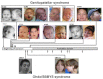
Facial features. Individuals with GPS can have:
Prominent cheeks
Nose with either a bulbous end or a broad or prominent base
Micro/retrognathia or prognathism
Bitemporal narrowing
Prominent eyes
See Figure 1.
Other features
Some children with GPS have hypotonia and/or laryngomalacia, which can exacerbate the feeding difficulties and respiratory difficulties present in some infants.
A minority of patients have delayed eruption of normal teeth, hypothyroidism, and mild to severe bilateral non-progressive sensorineural hearing loss.
Small bowel malrotation was seen in two affected individuals [Brugha et al 2011, Campeau et al 2012a] and duodenal rupture in another [Sankararaman et al 2012].
Other rarer features present in GPS are discussed in Campeau et al [2012a] and Penttinen et al [2009].
SBBYSS
Skeletal features. Most individuals have long thumbs and great toes. Some have patellar anomalies similar to those seen in GPS (i.e. aplasia or hypoplasia). Pectus chest deformity is seen in some. In one study, most patients had joint hypermobility in the upper extremities and large-joint stiffness in the lower extremities, sometimes to the point of contractures [Day et al 2008]. If not corrected, contractures can limit mobility.
Neurologic features. Patients have intellectual disability and severe, global developmental delay. In a study by Day et al [2008], children sat at 18 months and walked at 3.5 years. Fine motor skills and coordination were impaired. A minority were toilet trained and all required help dressing. A few children spoke at age 4.5 years, eventually making short sentences; receptive language was better than expressive language. Others communicated by vocalization only.
Some neonates have hypotonia, with feeding and respiratory difficulties requiring hospitalization.
Many have a smaller than average OFC but not frank microcephaly.
Genital anomalies. Cryptorchidism is seen in males. Females do not have genital anomalies. Pubertal delays are not seen.
Cardiac defects. Congenital heart defects are noted in about 50%. The most frequent defects are atrial septal defects, ventricular septal defects, and patent foramen ovale.
Facial features. Facial appearance is distinctive with a mask-like facies, blepharophimosis, and ptosis. Some have prominent cheeks, and a nose with either a bulbous end or a broad or prominent base. See Figure 1.
Other features
Feeding difficulties are seen. Contributing factors may be hypotonia and cleft palate, seen in one third of affected individuals [Clayton-Smith et al 2011].
Myopia and amblyopia are common. Some have lacrimal duct abnormalities.
Dental anomalies include natal teeth, retained primary dentition, and delayed eruption of teeth. Teeth can be misshapen or discolored.
A minority of patients have hypothyroidism, some of them having thyroid agenesis or hypoplasia.
Hearing loss, both conductive and sensorineural is often present.
Rarer features present in SBBYSS are discussed in [Day et al 2008].
Genotype-Phenotype Correlations
GPS. Known mutations causing GPS predict protein truncation and cluster in KAT6B exon 18, the last exon. Because mutations that predict truncation (nonsense, frameshift) that occur in the final exon of a gene typically do not result in nonsense-mediated decay of the mRNA, it is predicted that these KAT6B mutant alleles produce truncated proteins.
SBBYSS. Known mutations causing SBBYSS occur mostly in exon 18 but some individuals – often with an atypical SBBYSS phenotype – were found to have a mutation elsewhere in the gene. Nonsense/frameshift mutations in exons other than 18 are predicted to lead to nonsense-mediated decay and lack of protein expression from that allele. Mutation in KAT6B exon 18 typically occurs more distally than GPS-associated mutations (Figure 1).
Penetrance
KAT6B alleles with truncating mutations have a high penetrance. Truncating variants are not found in polymorphism databases and all family members with truncating variants reported have findings of GPS or SBBYSS [Campeau et al 2012a, Simpson et al 2012].
Nomenclature
GPS. The term genitopatellar syndrome was coined by Valérie Cormier-Daire [Cormier-Daire et al 2000].
SBBYSS. Dr. Ohdo first described a family in which children had blepharophimosis, ptosis, congenital heart defects, intellectual disability, and hypoplastic teeth [Ohdo et al 1986].
Subsequently, the Young-Simpson syndrome was described [Young & Simpson 1987]. Later the Young-Simpson syndrome was renamed the Say-Barber-Biesecker-Young-Simpson (SBBYS) variant of Ohdo syndrome (SBBYSS) [Say & Barber 1987, Biesecker 1991], which is discussed in this GeneReview chapter.
Of note, the disorder described by Dr. Ohdo was distinct from the SBBYS variant of Ohdo syndrome because the facial features differed, proteinuria was present, and skeletal anomalies were absent; furthermore, the mode of inheritance appeared to be autosomal recessive, autosomal dominant with reduced penetrance, or multifactorial.
Prevalence
The prevalence of KAT6B-related conditions is not known, but is estimated at less than one in a million individuals. Approximately 20 persons with GPS, and a similar number with SBBYSS, have been described in the literature.
Differential Diagnosis
For current information on availability of genetic testing for disorders included in this section, see GeneTests Laboratory Directory. —ED.
A minority of children clinically diagnosed as GPS or SBBYSS do not have mutations in KAT6B [Clayton-Smith et al 2011, Simpson et al 2012].
Other syndromes with patellar anomalies
Nail patella syndrome (LMX1B, autosomal dominant). Clinical tetrad of changes in the nails, knees, and elbows, and the presence of iliac horns
Small patella syndrome (TBX4, autosomal dominant). Aplasia or hypoplasia of the patellae and absent, delayed, or irregular ossification of the ischiopubic junctions or infraacetabular axe-cut notches
RAPADILINO syndrome (RECQL4, autosomal recessive). Irregular pigmentation with café au lait spots, small stature, palate defects, radial ray defects, patellar hypoplasia, and gastrointestinal abnormalities (See Baller-Gerold Syndrome, Genetically Related Disorders.)
Meier-Gorlin syndrome (pre-replication complex genes: ORC1, ORC4, ORC6, CDT1, CDC6, autosomal recessive). Severe intrauterine and postnatal growth retardation, microcephaly, bilateral microtia, and aplasia or hypoplasia of the patellae
Other syndromes with blepharophimosis
Blepharophimosis, ptosis, epicanthus inversus syndrome (BPES) (FOXL2, autosomal dominant). Reduced horizontal diameter of the palpebral fissures, ptosis, and a fold of skin that runs from the lower lids inwards and upwards (epicanthus inversus). Type II is isolated; type I is associated with premature ovarian insufficiency (POI).
Dubowitz syndrome (NSUN2 in one family [Martinez et al 2012], autosomal recessive). Blepharophimosis, ptosis, microcephaly, intellectual disability, growth retardation, eczema (features variably present) [Tsukahara & Opitz 1996]
Fetal alcohol spectrum disorders (FASD). Classic fetal alcohol syndrome includes in utero alcohol exposure, short palpebral fissures, flat midface, long and smooth philtrum, thin vermilion of the upper lip, pre- and post-natal growth retardation, microcephaly, and intellectual disability. Various degrees and combinations of these features, along with a variety of birth defects, may be present in individuals with FASD.
Other syndromes with agenesis of the corpus callosum and genital anomalies
Mowat-Wilson syndrome (ZEB2, autosomal dominant). Agenesis of the corpus callosum, intellectual disability, microcephaly, seizures, Hirschsprung disease, congenital heart disease, hypospadias, genitourinary anomalies, and short stature
X-linked lissencephaly with absent corpus callosum and ambiguous genitalia (ARX)
Other syndromes with agenesis of the corpus callosum and other malformations [Dobyns 1996]
Aicardi syndrome (X-linked dominant, gene unknown)
Hereditary motor and sensory neuropathy with agenesis of the corpus callosum (Andermann syndrome) (SLC12A6, autosomal recessive)
L1 syndrome (L1CAM, X-linked recessive)
Acrocallosal syndrome (KIF7, autosomal recessive)
Septooptic dysplasia (in some cases autosomal recessive with HESX1 mutations)
Other syndromes with genital anomalies and multiple congenital anomalies are reviewed in Neri & Opitz [1999], Simpson [1999].
Other syndromes that present with congenital contractures
Congenital contractural arachnodactyly (FBN2, autosomal dominant)
Cerebrooculofacioskeletal syndrome (COFS) (ERCC6 in some, autosomal recessive): progressive neurodegenerative disorder characterized by microcephaly, congenital cataracts, severe intellectual disability, facial dysmorphism, and arthrogryposis.
Amyoplasia
An approach to diagnosis of these syndromes is discussed elsewhere [Rink 2011].
Note to clinicians: For a patient-specific ‘simultaneous consult’ related to this disorder, go to  , an interactive diagnostic decision support software tool that provides differential diagnoses based on patient findings (registration or institutional access required).
Management
Evaluations Following Initial Diagnosis
To establish the extent of disease and needs of an individual diagnosed with KAT6B-related disorders, the following are recommended:
GPS and SBBYSS
Medical genetics consultation
Evaluation by developmental specialist
Feeding evaluation
Baseline hearing evaluation
Thyroid function tests
GPS
Evaluation of males for cryptorchidism
Orthopedic evaluation if contractures are present or feet/ankles are malpositioned
Hip radiographs to evaluate for femoral head dislocation
Renal ultrasound examination for hydronephrosis and cysts
Echocardiogram for congenital heart defects
Evaluation for laryngomalacia if respiratory issues are present
Evaluation by gastroenterologist as needed, particularly if bowel malrotation is suspected
SBBYSS
Ophthalmologic evaluation for lacrimal duct abnormalities, amblyopia, and other issues
Treatment of Manifestations
The following are appropriate:
Developmental. Educational intervention and speech therapy beginning in infancy because of the high risk for motor, cognitive, speech, and language delay
Skeletal features
Referral to an orthopedist for consideration of surgical release of contractures
Early referral to physical therapy to help increase joint mobility
Genital anomalies. Orchiopexy in males with undescended testes
Anal anomalies. Surgery as required
Renal anomalies. Referral to an urologist or nephrologist as indicated
Heart defects. Routine management
Facial features. In SBBYSS, ptosis surgery and cleft palate repair if present
Other features
Hearing aids as needed for hearing loss
Thyroid hormone replacement as needed
Intervention for respiratory and feeding issues as needed in the neonatal period
Referral to a gastroenterologist for evaluation and treatment of feeding difficulties and bowel malrotation
Surveillance
The following should be assessed annually:
Developmental progress
Ophthalmologic conditions such as amblyopia (which develops in a majority of patients with SBBYSS)
Thyroid function
Cardiac function if malformations are present
Renal function if hydronephrosis or multiple renal cysts are present
Contractures and/or scoliosis
Evaluation of Relatives at Risk
See Genetic Counseling for issues related to testing of at-risk relatives for genetic counseling purposes.
Therapies Under Investigation
Search ClinicalTrials.gov for access to information on clinical studies for a wide range of diseases and conditions. Note: There may not be clinical trials for this disorder.
Other
Genetics clinics, staffed by genetics professionals, provide information for individuals and families regarding the natural history, treatment, mode of inheritance, and genetic risks to other family members as well as information about available consumer-oriented resources. See the GeneTests Clinic Directory.
Genetic Counseling
Genetic counseling is the process of providing individuals and families with information on the nature, inheritance, and implications of genetic disorders to help them make informed medical and personal decisions. The following section deals with genetic risk assessment and the use of family history and genetic testing to clarify genetic status for family members. This section is not meant to address all personal, cultural, or ethical issues that individuals may face or to substitute for consultation with a genetics professional. To find a genetics or prenatal diagnosis clinic, see the GeneTests Clinic Directory.
Mode of Inheritance
KAT6B-related disorders are inherited in an autosomal dominant manner.
Risk to Family Members
Parents of a proband
To date, all individuals diagnosed with a KAT6B-related disorder have had a de novo mutation. Of note, one woman with relatively mild SBBYSS had a child with classic SBBYSS [Mhanni et al 1998]; however, to the authors’ knowledge molecular testing has not been performed in this family.
When the disease-causing mutation found in the proband cannot be detected in leukocyte DNA of either parent, two possible explanations are germline mosaicism in a parent or a de novo mutation in the proband. Although no instances of germline mosaicism have been reported, it remains a possibility.
Recommendations for the evaluation of parents of a proband with an apparent de novo mutation include molecular genetic testing of the parent if the KAT6B mutation has been identified in the proband.
Sibs of a proband
The risk to the sibs of the proband depends on the genetic status of the proband’s parents.
If the disease-causing mutation found in the proband cannot be detected in the leukocyte DNA of either parent, the risk to sibs is low but greater than that of the general population because of the possibility of germline mosaicism.
If a parent of the proband has germline mosaicism for a KAT6B mutation, the risk to the sibs of inheriting the mutation may be as high as 50%.
Offspring of a proband. To date, individuals with KAT6B-related disorders rarely reproduce. One woman with relatively mild SBBYSS had a child with classic SBBYSS [Mhanni et al 1998]; however, to the authors’ knowledge molecular testing has not been performed in this family.
Other family members. Because KAT6B-related disorders usually occur as the result of a de novo mutation, the risk to other family members is usually not increased.
Related Genetic Counseling Issues
Considerations in families with an apparent de novo mutation. When neither parent of a proband with an autosomal dominant condition has clinical evidence of the disorder it is likely that the proband has a de novo mutation.
Family planning
The optimal time for determination of genetic risk and discussion of the availability of prenatal testing is before pregnancy.
It is appropriate to offer genetic counseling (including discussion of potential risks to offspring and reproductive options) to couples who have had a child with a KAT6B-related disorder.
DNA banking is the storage of DNA (typically extracted from white blood cells) for possible future use. Because it is likely that testing methodology and our understanding of genes, mutations, and diseases will improve in the future, consideration should be given to banking DNA of affected individuals. See for a list of laboratories offering DNA banking.
Prenatal Testing
Prenatal diagnosis for pregnancies at increased risk is possible by analysis of DNA extracted from fetal cells obtained by amniocentesis usually performed at approximately 15 to 18 weeks’ gestation or chorionic villus sampling (CVS) at approximately ten to 12 weeks’ gestation. The disease-causing mutation of an affected family member must have been identified in the family before prenatal testing can be performed.
Note: Gestational age is expressed as menstrual weeks calculated either from the first day of the last normal menstrual period or by ultrasound measurements.
Preimplantation genetic diagnosis (PGD) may be available for families in which the disease-causing mutation has been identified. For laboratories offering PGD, see .
Note: It is the policy of GeneReviews to include in GeneReviews™ chapters any clinical uses of testing available from laboratories listed in the GeneTests™ Laboratory Directory; inclusion does not necessarily reflect the endorsement of such uses by the author(s), editor(s), or reviewer(s).
Molecular Genetics
Information in the Molecular Genetics and OMIM tables may differ from that elsewhere in the GeneReview: tables may contain more recent information. —ED.
Table A. KAT6B-Related Disorders: Genes and Databases
| Gene Symbol | Chromosomal Locus | Protein Name | Locus Specific | HGMD |
|---|---|---|---|---|
| KAT6B | 10q22 | Histone acetyltransferase KAT6B | KAT6B@LOVD | MYST4 |
Table B. OMIM Entries for KAT6B-Related Disorders (View All in OMIM)
Normal allelic variants. KAT6B comprises 18 exons, the first two of which are untranslated. Three protein isoforms exist owing to alternative splice donor sites used for exon 8. NM_012330.3 represents the longest transcript and encodes the longest isoform. Normal coding variants present in at least 1% of the population are in Table 4.
Pathologic allelic variants. All pathologic variants known to date are either nonsense or frameshift mutations that cause a premature truncation of the protein. All variants were de novo. Variants occurring in more than one individual are shown in Table 4.
GPS-causing mutations are in the proximal portion of the last exon (exon 18) (Figure 1) and lead to the expression of a protein without a C-terminal portion (see Normal gene product).
SBBYSS-causing mutations occur most often in the distal portion of exon 18 (the last exon) or exceptionally in earlier exons, and presumably lead to nonsense-mediated decay and no protein production.
Table 4. Selected KAT6B Allelic Variants
| Class of Variant Allele | DNA Nucleotide Change | Protein Amino Acid Change | Reference Sequences |
|---|---|---|---|
| Normal | c.3649G>T | p.Ala1217Ser (rs57372986) 1 | NM_012330 NP_036462 |
| c.4495G>A | p.Val1499Ile (rs374032) 1 | ||
| c.4835G>A | p.Arg1612His (rs72803461) 1 | ||
| Pathologic - GPS | c.3769_3772delTCTA | p.Lys1258GlyfsX13 | |
| Pathologic -SBBYSS | c.4205_4206del | p.Ser1402CysfsX5 | |
| c.5201_5210dup | p.Gln1737HisfsX41 |
See Quick Reference for an explanation of nomenclature. GeneReviews follows the standard naming conventions of the Human Genome Variation Society (www
.hgvs.org). 1. Description in dbSNP, a database of single nucleotide polymorphisms (SNPs) and multiple small-scale variations that include insertions/deletions, microsatellites, and non-polymorphic variants.
Normal gene product. KAT6B, a ubiquitously expressed histone acetyltransferase, regulates the expression of various genes, and preferentially acetylates lysine 14 of histone H3 in an in vitro assay [Doyon et al 2006]. KAT6B interacts with the proteins RUNX2, BRPF1, ING5, PPAR-alpha, and atrophin-1; however, the precise roles of these proteins in the mechanism of action of KAT6B are not completely understood (reviewed by Yang & Ullah [2007]).
Abnormal gene product. It is possible that haploinsufficiency or loss of a function mediated by the C-terminal region causes the features common to both conditions, whereas gain-of-function activities would explain the features unique to GPS (such as anomalies of the spine, ribs, and pelvis; renal cysts; hydronephrosis; and agenesis of the corpus callosum). Since only some of the binding partners of KAT6B are known, it is difficult to hypothesize which of these are involved in the pathogenesis of KAT6B-related diseases.
Resources
No specific resources for KAT6B-Related Disorders have been identified by GeneReviews staff.
References
Medical Genetic Searches: A specialized PubMed search designed for clinicians that is located on the PubMed Clinical Queries page 
Literature Cited
- Biesecker LG. The Ohdo blepharophimosis syndrome: a third case. J Med Genet. 1991;28:131–4. [PMC free article: PMC1016784] [PubMed: 2002485]
- Brugha R, Kinali M, Aminu K, Bridges N, Holder SE. Genitopatellar syndrome: a further case. Clin Dysmorphol. 2011;20:163–5. [PubMed: 21412151]
- Campeau PM, Kim JC, Lu JT, Schwartzentruber JA, Abdul-Rahman OA, Schlaubitz S, Murdock DM, Jiang MM, Lammer EJ, Enns GM, Rhead WJ, Rowland J, Robertson SP, Cormier-Daire V, Bainbridge MN, Yang XJ, Gingras MC, Gibbs RA, Rosenblatt DS, Majewski J, Lee BH. Mutations in KAT6B, encoding a histone acetyltransferase, cause Genitopatellar syndrome. Am J Hum Genet. 2012;90:282–9. [PMC free article: PMC3276659] [PubMed: 22265014]
- Campeau PM, Lu JT, Dawson BC, Fokkema IF, Robertson SP, Gibbs RA, Lee BH. The KAT6B-related disorders Genitopatellar syndrome and Ohdo/SBBYS syndrome have distinct clinical features reflecting distinct molecular mechanisms. Hum Mutat. 2012;33:1520–5. [PubMed: 22715153]
- Clayton-Smith J, O'Sullivan J, Daly S, Bhaskar S, Day R, Anderson B, Voss AK, Thomas T, Biesecker LG, Smith P, Fryer A, Chandler KE, Kerr B, Tassabehji M, Lynch SA, Krajewska-Walasek M, McKee S, Smith J, Sweeney E, Mansour S, Mohammed S, Donnai D, Black G. Whole-exome-sequencing identifies mutations in histone acetyltransferase gene KAT6B in individuals with the Say-Barber-Biesecker variant of Ohdo syndrome. Am J Hum Genet. 2011;89:675–81. [PMC free article: PMC3213399] [PubMed: 22077973]
- Cormier-Daire V, Chauvet ML, Lyonnet S, Briard ML, Munnich A, Le Merrer M. Genitopatellar syndrome: a new condition comprising absent patellae, scrotal hypoplasia, renal anomalies, facial dysmorphism, and mental retardation. Med Genet. 2000;37:520–4.
- Day R, Beckett B, Donnai D, Fryer A, Heidenblad M, Howard P, Kerr B, Mansour S, Maye U, McKee S, Mohammed S, Sweeney E, Tassabehji M, de Vries BB, Clayton-Smith J. A clinical and genetic study of the Say/Barber/Biesecker/Young-Simpson type of Ohdo syndrome. Clin Genet. 2008;74:434–44. [PubMed: 18798845]
- Dobyns WB. Absence makes the search grow longer. Am J Hum Genet. 1996;58:7–16. [PMC free article: PMC1914936] [PubMed: 8554070]
- Doyon Y, Cayrou C, Ullah M, Landry AJ, Côté V, Selleck W, Lane WS, Tan S, Yang XJ, Côté J. ING tumor suppressor proteins are critical regulators of chromatin acetylation required for genome expression and perpetuation. Mol Cell. 2006;21:51–64. [PubMed: 16387653]
- Kraft M, Cirstea IC, Voss AK, Thomas T, Goehring I, Sheikh BN, Gordon L, Scott H, Smyth GK, Ahmadian MR, Trautmann U, Zenker M, Tartaglia M, Ekici A, Reis A, Dörr HG, Rauch A, Thiel CT. Disruption of the histone acetyltransferase MYST4 leads to a Noonan syndrome-like phenotype and hyperactivated MAPK signaling in humans and mice. J Clin Invest. 2011;121:3479–91. [PMC free article: PMC3163944] [PubMed: 21804188]
- Mhanni AA, Dawson AJ, Chudley AE. Vertical transmission of the Ohdo blepharophimosis syndrome. Am J Med Genet. 1998;77:144–8. [PubMed: 9605288]
- Martinez FJ, Lee JH, Lee JE, Blanco S, Nickerson E, Gabriel S, Frye M, Al-Gazali L, Gleeson JG. Whole exome sequencing identifies a splicing mutation in NSUN2 as a cause of a Dubowitz-like syndrome. J Med Genet. 2012;49:380–5. [PubMed: 22577224]
- Neri G, Opitz J. Syndromal (and nonsyndromal) forms of male pseudohermaphroditism. Am J Med Genet. 1999;89:201–9. [PubMed: 10727995]
- Ohdo S, Madokoro H, Sonoda T, Hayakawa K. Mental retardation associated with congenital heart disease, blepharophimosis, blepharoptosis, and hypoplastic teeth. J Med Genet. 1986;23:242–4. [PMC free article: PMC1049635] [PubMed: 3723552]
- Penttinen M, Koillinen H, Niinikoski H, Mäkitie O, Hietala M. Genitopatellar syndrome in an adolescent female with severe osteoporosis and endocrine abnormalities. Am J Med Genet A. 2009;149A:451–5. [PubMed: 19208376]
- Rink BD. Arthrogryposis: a review and approach to prenatal diagnosis. Obstet Gynecol Surv. 2011;66:369–77. [PubMed: 21851751]
- Sankararaman S, Kurepa D, Velayuthan S, Kakkilaya V, Gates T, Ursin S, Chen H. Another case of genitopatellar syndrome: a case report with additional rare coexistences. Clin Dysmorphol. 2012;21:226–8. [PubMed: 22922314]
- Say B, Barber N. Mental retardation with blepharophimosis. J Med Genet. 1987;24:511. [PMC free article: PMC1050218] [PubMed: 3656379]
- Simpson JL. Genetics of the female reproductive ducts. Am J Med Genet. 1999;89:224–39. [PubMed: 10727998]
- Simpson MA, Deshpande C, Dafou D, Vissers LE, Woollard WJ, Holder SE, Gillessen-Kaesbach G, Derks R, White SM, Cohen-Snuijf R, Kant SG, Hoefsloot LH, Reardon W, Brunner HG, Bongers EM, Trembath RC. De novo mutations of the gene encoding the histone acetyltransferase KAT6B cause Genitopatellar syndrome. Am J Hum Genet. 2012;90:290–4. [PMC free article: PMC3276665] [PubMed: 22265017]
- Tsukahara M, Opitz JM. Dubowitz syndrome: review of 141 cases including 36 previously unreported patients. Am J Med Genet. 1996;63:277–89. [PubMed: 8723121]
- White SM, Adès LC, Amor D, Liebelt J, Bankier A, Baker E, Wilson M, Savarirayan R. Two further cases of Ohdo syndrome delineate the phenotypic variability of the condition. Clin Dysmorphol. 2003;12:109–13. [PubMed: 12868473]
- Yang XJ, Ullah M. MOZ and MORF, two large MYSTic HATs in normal and cancer stem cells. Oncogene. 2007;26:5408–19. [PubMed: 17694082]
- Young ID, Simpson K. Unknown syndrome: abnormal facies, congenital heart defects, hypothyroidism, and severe retardation. J Med Genet. 1987;24:715–6. [PMC free article: PMC1050356] [PubMed: 3430551]
Suggested Reading
- Fokkema IF, Taschner PE, Schaafsma GC, Celli J, Laros JF, den Dunnen JT. LOVD v.2.0: the next generation in gene variant databases. Hum Mutat. 2011;32:557–63. [PubMed: 21520333]
Chapter Notes
Author Notes
Dr. Brendan Lee’s Web site
Dr. Philippe Campeau’s Web site
About the Authors’ research. The spectrum of our research program extends from gene identification in human disease, to correlating mechanisms of disease with normal biologic processes, to measuring and manipulating these pathways for diagnosis and treatment in humans and in animal models.
Revision History
10 January 2013 (cd) Revision: sequence analysis and prenatal diagnosis available clinically
13 December 2012 (me) Review posted live
19 June 2012 (pc/bl) Original submission

GeneReviews™ [Internet].
Pagon RA, Bird TD, Dolan CR, et al., editors.
Seattle (WA): University of Washington, Seattle; 1993-.
-
The KAT6B-related disorders Genitopatellar syndrome and Ohdo/SBBYS syndrome have distinct clinical features reflecting distinct molecular mechanisms.
[Hum Mutat. 2012]
The KAT6B-related disorders Genitopatellar syndrome and Ohdo/SBBYS syndrome have distinct clinical features reflecting distinct molecular mechanisms.Campeau PM, Lu JT, Dawson BC, Fokkema IF, Robertson SP, Gibbs RA, Lee BH. Hum Mutat. 2012 Nov; 33(11):1520-5. Epub 2012 Jul 12.
-
Whole-exome-sequencing identifies mutations in histone acetyltransferase gene KAT6B in individuals with the Say-Barber-Biesecker variant of Ohdo syndrome.
[Am J Hum Genet. 2011]
Whole-exome-sequencing identifies mutations in histone acetyltransferase gene KAT6B in individuals with the Say-Barber-Biesecker variant of Ohdo syndrome.Clayton-Smith J, O'Sullivan J, Daly S, Bhaskar S, Day R, Anderson B, Voss AK, Thomas T, Biesecker LG, Smith P, et al. Am J Hum Genet. 2011 Nov 11; 89(5):675-81.
-
Mutations in KAT6B, encoding a histone acetyltransferase, cause Genitopatellar syndrome.
[Am J Hum Genet. 2012]
Mutations in KAT6B, encoding a histone acetyltransferase, cause Genitopatellar syndrome.Campeau PM, Kim JC, Lu JT, Schwartzentruber JA, Abdul-Rahman OA, Schlaubitz S, Murdock DM, Jiang MM, Lammer EJ, Enns GM, et al. Am J Hum Genet. 2012 Feb 10; 90(2):282-9. Epub 2012 Jan 19.
-
KANSL1-Related Intellectual Disability Syndrome
[GeneReviews™. 1993]
KANSL1-Related Intellectual Disability SyndromeKoolen DA, de Vries BBA. GeneReviews™. 1993
-
Review Vertical transmission of the Ohdo blepharophimosis syndrome.
[Am J Med Genet. 1998]
Review Vertical transmission of the Ohdo blepharophimosis syndrome.Mhanni AA, Dawson AJ, Chudley AE. Am J Med Genet. 1998 May 1; 77(2):144-8.
-
KAT6B-Related Disorders - GeneReviews™
KAT6B-Related Disorders - GeneReviews™Bookshelf
-
Journey to the patient-centered medical home: a qualitative analysis of the expe...
Journey to the patient-centered medical home: a qualitative analysis of the experiences of practices in the National Demonstration Project.Ann Fam Med. 2010 ;8 Suppl 1:S45-56; S92. doi: 10.1370/afm.1075.PubMed
-
Joint working in community mental health teams: implementation of an integrated ...
Joint working in community mental health teams: implementation of an integrated care pathway.Health Soc Care Community. 2004 Nov ;12(6):527-36.PubMed
-
Identifiers, or "red flags," of complexity and need for integrated care.
Identifiers, or "red flags," of complexity and need for integrated care.Med Clin North Am. 2006 Jul ;90(4):703-12.PubMed
-
Identification of somatic and anxiety symptoms which contribute to the detection...
Identification of somatic and anxiety symptoms which contribute to the detection of depression in primary health care.Eur Psychiatry. 2004 Aug ;19(5):250-7.PubMed
Your browsing activity is empty.
Activity recording is turned off.
See more...