|
Two new studies appear to highlight the role of glial cells—the nervous system's equivalents to the body's immune cells—in methamphetamine abuse.
By Patick Zickler, NIDA NOTES Contributing Writer
NIDA-supported researchers have produced brain images demonstrating that structures in an area called the striatum expand in volume during early methamphetamine abuse, then regress toward normal. The investigators believe their findings likely are attributable to neuroprotective cells in the brain mounting an initial attempt to counteract the drug's toxic effects, which continued exposure subsequently overwhelms. In a related result, scientists working with mice have produced evidence that methamphetamine may prompt cells that normally serve neuroprotective functions to instead attack healthy brain cells.
STRUCTURAL FLUCTUATION SUGGESTS GLIAL ACTIVATION
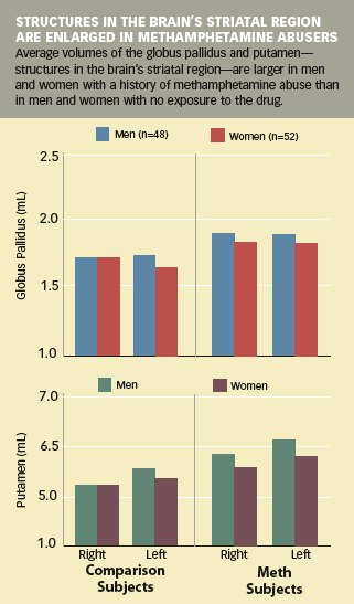
Dr. Linda Chang (now at the University of Hawaii) and colleagues at the University of California, Los Angeles, used magnetic resonance imaging to measure the volumes of striatal brain structures, including the putamen and globus pallidus, in a group of methamphetamine abusers. The studied individuals, 26 women and 24 men (average age 31 years), had abused methamphetamine (average 1.6 g/day on 6.3 days/week) for periods ranging from 4 to 15 years. All had been abstinent for periods ranging from 1 week to 4 years at the time of the study; 44 also took tests of verbal memory and intelligence, gross and fine motor function, mood, executive function, and other capacities likely to be affected by striatal damage.
The researchers expected to find that the methamphetamine abusers' striatal regions were smaller than those of a comparison group of age- and gender-matched individuals with no history of methamphetamine abuse. Such a finding would be consistent with previous research showing that methamphetamine reduces the density of striatal dopamine terminals. Instead, says Dr. Chang, "Contrary to our hypothesis, striatal volumes were larger in the methamphetamine abusers as a group." The size difference was greatest among individuals with less cumulative exposure to the drug, and smaller among those with more. Those with the most exposure also performed slightly worse on neuropsychological tests of verbal fluency and visual-motor coordination.
Dr. Chang believes the surprising increase in striatal volumes of methamphetamine abusers may reflect the activity of glia—cells that provide protective and reparative functions for the brain's main functional cells, the neurons. When molecules potentially harmful to neurons penetrate the brain, glia mount a response resembling the inflammation and scar tissue formation associated with immune responses in other parts of the body. Possibly, Dr. Chang suggests, methamphetamine provokes glia to react in this way, leading to an increase in regional volume analogous to the swelling seen in bodily immune responses. Subsequently, she speculates, the glial response may taper off as cumulative exposure to the drug—and neuron damage—mount. Continued abuse results in damage that is manifested in decreased cognitive performance.
"This work is consistent with an increasing body of research that shows a relationship between methamphetamine exposure and structural changes in the brain," says Dr. Steven Grant of NIDA's Division of Clinical Neuroscience and Behavioral Research. "It links methamphetamine abuse, structural change, and functional deficits and suggests that the magnitude of these effects is related to the degree of abuse. We don't understand what is happening at deeper levels, but the observations made in this study suggest that the volume changes are related to methamphetamine's direct or indirect effect on glial cells. We still need to understand how structural changes result in functional deficits; how much, if any, of this damage can be reversed; and how methamphetamine acts at the cellular level."
IN MICE, METHAMPHETAMINE MISDIRECTS GLIA TO ATTACK BRAIN CELLS
A study by Drs. Donald Kuhn and David Thomas and colleagues at Wayne State University School of Medicine indicates that methamphetamine's toxic effects may include subverting some glial cells to attack rather than preserve neurons. Specifically, their results indicate that the drug incites a subset of glia called microglia to mount an immune response against dopamine neurons.
Normally, microglia protect neurons against toxic injury by several mechanisms. They detect and bind to invading molecules, including viruses or bacteria, making them easily accessible to destructive immune system cells such as lymphocytes. As well, they produce compounds, some toxic, to help contain or eliminate the danger. Methamphetamine, the new study suggests, causes dopamine neurons to release a signal that decoys the microglia into turning these normally protective responses against the neurons themselves. When that happens, Dr. Kuhn says, "The microglia aren't reacting to methamphetamine's neural damage. Instead, they are active participants in the drug's neurotoxicity."
To begin their experiments, the researchers reasoned that if microglia contribute to methamphetamine toxicity to dopamine terminals, compounds that protect against such toxicity might do so, at least in part, by inhibiting microglial activation. Their first hypothesis, accordingly, was that the compound MK-801, which is known to be protective, blunts microglial activation. The team showed this to be the case by exposing cell cultures of mouse microglia to two proteins known to precipitate damaging microglial responses: lipopolysaccharide (LPS) and HIV Tat, a derivative of the human immunodeficiency virus. Compared with LPS and HIV Tat exposure without pretreatment, exposure following pretreatment with MK-801 significantly reduced the amount of two protein products of microglial activation, called cyclooxygenase-2 (Cox-2) and tumor necrosis factor-α (TNF-α). Dextromethorphan (DXM), a compound biochemically similar to MK-801, had the same effect.
"These results suggested that both MK-801 and dextromethorphan exert direct action on the microglial cells in culture to block the activation process," Dr. Kuhn says. Having determined that the two compounds block microglial activation in vitro, the researchers next hypothesized that they would also do so in living animals.
Drs. Kuhn and Thomas injected mice with either MK-801 or DXM and then methamphetamine (5 mg/kg of body weight) 15 minutes later, repeating this sequence four times at 2-hour intervals. A control group of mice received the same regimen, but with saline substituted for methamphetamine. Forty-eight hours after the last injection, the researchers assayed the brains of the mice for Cox-2 and TNF-α, the indicators of microglia activation, and for striatal dopamine levels, a widely used index of damage to dopamine neurons. Dr. Kuhn says, "We found that both DXM and MK-801 significantly reduced the markers of striatal microglial activation associated with methamphetamine exposure and protected against dopamine nerve terminal damage in the striatum. The close association between the ability of MK-801 and DXM to significantly lower both microglial activation and neuronal damage suggests a causal link between the two. It looks as though the damage associated with methamphetamine abuse is the result of microglial action."
The apparent association of microglia and damage to dopamine neurons has implications beyond what it may reveal about methamphetamine abuse, says Dr. Jerry Frankenheim of NIDA's Division of Basic Neuroscience and Behavioral Research. "Microglia are the primary immune defense cells in the brain. They safeguard neural functions, yet excessive activation can cause microglia to harm neurons. Other research implicates microglial involvement in a wide range of neurodegenerative disorders, including Alzheimer's disease, Parkinson's disease, and stroke. Understanding how methamphetamine is able to decoy microglia into a destructive rather than reparative role could also help explain the processes involved in these other disorders."
SOURCES
Chang, L., et al. Enlarged striatum in abstinent methamphetamine abusers: A possible compensatory response. Biological Psychiatry 57(9):967-974, 2005. [Abstract]
Thomas, D.M., and Kuhn, D.M. MK-801 and dextromethorphan block microglial activation and protect against methamphetamine-induced neurotoxicity. Brain Research 1050(1-2):190-198, 2005. [Abstract]
Volume 20, Number 6 (July 2006)
|
|
|