Visual Function Core (VFC)
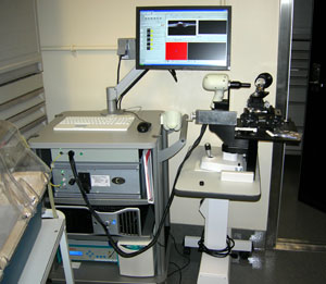
Optical coherence tomography machine used to provide and overview of the retina's structure.
The NEI Visual Function Core provides expertise in using non-invasive techniques to measure visual function in animal models. Currently, the core provides training to use electrophysiological and imaging techniques and access to evaluate eye function.
Electroretinograms (ERG) used by the VFC affords a quantitative, objective, and noninvasive method to examine light-evoked neuronal activity and is commonly used to study the functional integrity of normal and diseased retinas. A state-of-art Optical Coherence Tomography (OCT) machine is also available to generate high quality optical section of retina in live animals.
In the near future, behavioral tests of visual function and other techniques for evaluating animal visual performance will become available. Please contact core head for detail applications and service.
Staff
| Name | Title | |
|---|---|---|
| Haohua Qian, Ph.D. | Head | qianh@nei.nih.gov |
Last Reviewed: March 2011

