Histopathology Core Facility
Facilities and Equipment
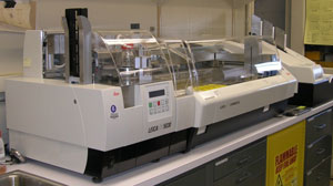
Coverslipper and multistainer for histology and cytopathology.
The National Eye Institute's Histopathology Core Facility provides expertise and up-to-date technical services for histological (under a light microscope or electron microscope) and cytological analyses for all of NEI and other NIH scientists. Additionally, the NEI Histopathology Core provides services for the pathological diagnosis of ocular diseases for clinicians. This data can be made available in a visual or quantitative format.
The NEI Histopathology Core Facility is a Clinical Laboratory Improvement Approved (CLIA) lab, CLIA ID #21D0952944, licensed by the U.S. Department of Health & Human Services.
Core Services
The Core staff provides technical assistance in the development and optimization of histopathological and morphometric techniques, including the following:
Sample preparation and sectioning
- Fixation of cells, tissues and organs
- Grossing tissues and organs, particularly the eyes (human and animal)
- Embedding frozen tissues or organs in OCT (optimal cutting temperature)
- Embedding samples in methacrylate or paraffin for light microscopy
- Embedding samples in resin for electron microscopy
- Sectioning frozen samples with a cryostat
- Sectioning of embedded samples with a microtome or ultramicrotome
Slide processing
- Routine histological stains such as H&E or PAS II and stains such as Gram stains, AFB, silver, etc.
- Immunostaining of human cells, tissues and organs with enzyme histochemistry (acid phosphatase, alkaline phosphatase, HRP, ABC avidin-biotin-complex)
- Microdissection and molecular analyses for research and patient diagnostic slides
Staff
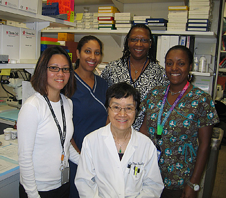
Histology Core: (left to right) Michelle Roof, Janelle Cole, Chi-Chao Chan (seated), Doretha Huntely , and Iris Wise.
| Name | Title | |
|---|---|---|
| Chi-Cho Chan, M.D. | Head | ccc@helix.nih.gov |
| Mones Abu-Asab | Biologist/Electron Microscopist | mones@nih.gov |
| Iris Wise | Lead Histologist | milleri@intra.nei.nih.gov |
| Janelle Coles | Histologist | colesjs@mail.nih.gov |
| Michelle Roof | Histologist (part time) | roofm@mail.nih.gov |
| Narva Thompson | Histologist (part time) | thompsonn2@mail.nih.gov |
Last Reviewed: November 2012

