|
A small study using a new imaging technique revealed an association between MDMA (Ecstasy) abuse and lower gray matter density in key brain structures that affect language, movement, and vital functions such as breathing and heartbeat.
| New Brain Imaging Technique Detects Brain Density Differences in MDMA Abusers |
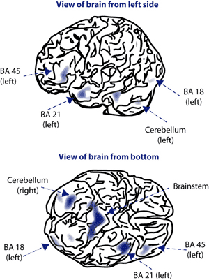 Preliminary results from voxel-based morphometry suggest
decreased density in some brain areas of people who abuse
MDMA. Affected areas included the bilateral Brodmann area (BA)
18, the left BA 21 and left BA 45, and the bilateral cerebellum and
midline brainstem. |
|
Drs. Ronald Cowan, Perry Renshaw, In Kyoon Lyoo, and colleagues at the McLean Hospital in Belmont, Massachusetts, set out to find evidence in people for an MDMA effect that some animal studies have pointed to: damage to nerve fibers that deliver the neurotransmitter serotonin to certain regions of the brain. Serotonin promotes the growth of neurons and glial cells, which together comprise the brain's gray matter. The researchers hypothesized, accordingly, that if the suspected effect were true, MDMA-abusing individuals would come to have less abundant gray matter in the regions served by the damaged fibers compared with nonabusers of the drug. Such hypotheses have recently become testable with the development of a new imaging technique, voxel-based morphometry (VBM), which measures gray matter density.
The researchers examined 60 volunteers aged 18 to 35 at McLean Hospital in Belmont, Massachusetts. Thirty-one of the study participants had abused MDMA at least 5 times, while 29 had never taken it. Brain images of the MDMA abusers were taken at least 3 weeks after their last exposure to the club drug. The results bore out the researchers' hypothesis: They showed smaller concentrations of gray matter in the MDMA abusers than in the controls. The reductions in density do not amount to "holes" in the brain, Dr. Cowan says, but are instead subtle but statistically significant differences seen in several brain regions. Some of the regions with less gray matter were parts of the neocortex that may be involved in word definition. Other implicated areas were the cerebellum, which controls movement, motor learning, and spatial sense, and the brainstem, which controls respiration and heart rhythms and integrates information between the peripheral nervous system and the spinal cord.
The study findings are consistent with the previously existing evidence that MDMA may damage serotonin fibers, but they do not prove it. Dr. Cowan cautions that his study had too few participants to fully rule out the possibility that the MDMA abusers had less gray matter before they took the drug.
Source
Cowan, R.L., et al. Reduced cortical gray matter density in human MDMA (ecstasy) users: A voxel-based morphometry study. Drug and Alcohol Dependence 72(3):225-235, 2003.
[Abstract]
Volume 19, Number 5 (January 2005)
|
|
|