Three-D Informatics Group
A Group Dedicated to Research and Education in:
- computer graphics
- image processing
- modeling, and
- machine and human perception
ITK: Insight Toolkit
The Insight Toolkit (ITK) project is developing a public, open-source software library of leading-edge algorithms for the segmentation (image partitioning) and registration (image alignment) of high-dimensional biomedical image data. ITK improvements are incorporating the latest advances in multi-core processors in laptop systems, larger memory capacity across all computing environments, ubiquitous graphics processing units (GPUs), and the increasing availability of consumer-grade parallel computing systems. ITK is being integrated into software across a wide array of scientific and medical disciplines, from image-guided surgery and tumor volume measurement to biological discovery through high-resolution electron microscopy. ITK support, development, and maintenance are provided through a university and industry consortium and NLM/LHNCBC/OHPCC intramural research staff.
High Resolution Electron Microscopy
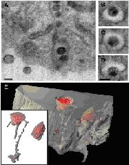
Beyond data collection, this group is engaged in aggressive image analysis programs in partnership with the High Resolution Microscopy Laboratory at the National Cancer Institute. 3D Informatics staff members are developing new techniques using the Insight Toolkit from the Visible Human Project to study sub-cellular structure from 3D dual-beam scanning electron micrographs and 3D transmission-electron tomograms of cultured cells exhibiting critical pathologies such as melanoma and HIV/AIDS. This effort includes analysis as well as visualization and rendering of complex microscopy data, integrating the HPCC facilities in 3D printing to support intramural, trans-NIH research.
Computational Photography Project for Pill Identification
In a national effort to promote patient safety, the National Library of Medicine (NLM) proposes to create a comprehensive, public digital image inventory of the nation's commercial prescription solid dose medications. The primary intention of this effort is create a test data collection for the advancement of automatic pharmaceutical identification through computer analysis from photographic data. NLM expects to promote computer-based image research applied to the domain of content-based information retrieval (CBIR) of solid-dose pharmaceuticals and anticipates the need for generating a test environment, including variations of photographs of the same drug or sample under different environments.
Immersive Display of 3-D Printing of X-ray CT Phantoms
We are seeking ground-truth 3D X-ray CT phantoms with gradations of Hounsfield units that are indistinguishable from scans of human subjects. We modified a 3D printer, a ZCorp Spectrum 510, adding an iodine-based contrast agent, and printed physical models using a powder which consists mainly of cellulose and cornstarch. We scanned these 3D models with a Siemens Somatom Definition AS 128-slice CT scanner. By adjusting the level of iodine within the model, we are able to achieve Hounsfield units as high as 1056, mimicking bone, and as low as -450, similar to pulmonary tissue. We demonstrate how to generate grayscale images within a 3D model that can be imaged using a CT scanner. Unlike solid tumor phantoms, these models can accurately mimic lesions with indistinct boundaries similar to metastatic disease.


