
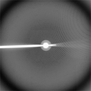
X-Ray diffraction image created from a crystal of Bacteriophage HK97.
Source: John Johnson, The Scripps Research Institute
This year marks the 100th anniversary of X-ray diffraction technology. Developed in 1912, this important tool enables researchers to figure out the 3-D structure of a molecule by beaming X-rays, often through its crystallized form. More than 85% of the protein structures we know today have been determined via this method.
For more information about x-ray diffraction, I recommend Structural Biology Fact Sheet and The Structures of Life: X-ray Crystallography.

Here you see the X-ray diffraction image that James Watson and Francis Crick used to decipher the double helix structure of DNA in 1953.
And now for a trivia question! As some of you may know, one of my hobbies is playing the guitar—a guitar that happens to have a DNA double helix inlaid on its fretboard. All special guitars should have a name. B.B. King has Lucille. Eric Clapton had Blackie. After which famous scientist, responsible for the image used by Watson and Crick, is my guitar named?


 Get new blog posts by email
Rosa? (Rosalind Franklin)
Rosalind Franklin, of course. Her name should be mentioned next to the picture. The sentence “Here you see the X-ray diffraction image that James Watson and Francis Crick used to decipher the double helix structure of DNA in 1953″ gives the impression as if Watson and Crick had produced this beautiful picture rather than Rosalind Franklin. It is well known now that Watson was given this picture without her knowledge.
Miral, well articulated. I’m surprised that Watson and Crick are still mentioned without also mentioning Rosalind Franklin.
I stand corrected. I see she was mentioned, in letters written upside down.
It’s amazing when you consider that Sir William Lawrence Bragg (1890-1971) was only 21 years old when he and his father successfully developed x-ray crystallography as a tool for structure elucidation. That early discovery and his extraordinary ability and influence destined DNA and protein structure elucidation history in Cambridge/London. Unfortunately, x-ray structure alone does not tell us that much about the function of all those proteins. Understanding function requires “new ways of thinking about them”. To quote WL Bragg, “The important thing in science is not so much to obtain new facts as to discover new ways of thinking about them”.
I’m surprised and dismayed that this note ignores the rich history of x-ray diffraction in fields other than medicine. The discovery and elucidation of x-ray diffraction was honored by the 1915 Nobel Prize in Physics; many pathbreaking advances in physics & cnemistry followed, setting the stage for the diffraction measurements by Franklin that were interpreted by Watson & Crick. This is but one of many instances in which advances in physics and chemistry directly enabled medical breakthroughs (CT Scans and MRI scans are prominent and recent examples). It’s unfortunate that Dr. Collins didn’t call attention to such interdisciplinary cross-fertilization.