Home » Photos, Images, and Videos » Search Results
Photos, Images, and Videos
Search Results
Instructions for Downloading Photographs and Images
 |
Description: Diabetic Retinopathy, Video Illustration. Diabetic eye disease refers to a group of eye problems that can occur as a result of diabetes. Without diagnosis and treatment, diabetic eye disease can cause severe vision loss or even blindness. Diabetic retinopathy is the most common diabetic eye disease and is a leading cause of blindness in persons with diabetes. It is caused by changes in the blood vessels of the retina. The retina is the light-sensitive tissue at the back of the eye. A healthy retina is necessary for good vision. In some people with diabetic retinopathy, blood vessels of the retina may swell and leak fluid or blood. In other people with diabetes, abnormal new blood vessels grow on the surface of the retina. At first, diabetic retinopathy may not cause any changes in vision. But over time, diabetic retinopathy can get worse. At first, a person might see spots floating in their vision or may notice a general blurring of vision. Eventually, diabetic retinopathy may cause vision loss and even blindness. Credit: National Eye Institute, National Institutes of Health View Animation (Quicktime) Ref#: VA07 |
|
 |
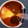
Description: Cataract, video illustration. A cataract is a clouding of the lens in your eye that affects vision. Most cataracts are related to aging. To understand how a cataract can affect your vision, it helps to understand a little about the structure of your eye. The lens is clear and lies between the iris and the pupil. It works much like a camera lens, focusing light, or an image, on the retina. The retina is the light-sensitive tissue at the back of the eye. Once the light or image reaches the retina, it is changed into nerve signals that are sent to the brain. Besides focusing light on the retina, the lens also adjusts the eye's focus, letting us see things clearly both up close and far away. The lens must be clear for the retina to receive a sharp image. The lens is made of mostly water and protein. The protein is arranged in a precise way that allows light to pass through it without distortion. But as we age, proteins in the lens clump together, causing a cataract. At first the protein may clump together and cloud in small area of the lens. Over time, the clumps may increase in size and number, clouding more of the lens. If the lens is cloudy from a cataract, the image you see will be blurred and colors may appear faded. Credit: National Eye Institute, National Institutes of Health View Animation (Quicktime) Ref#: VA08 |
|
 |
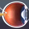
Description: Glaucoma, video illustration. A clear fluid flows through the anterior segment at the front of the eye nourishing tissues. At the open angle where the cornea and iris meet the fluid flows through a spongy meshwork and leaves the eye. But if it passes through too slowly pressure inside the eye rises to a level that damages the optic nerve resulting in vision loss from open-angle glaucoma. Untreated, the vision loss progresses from normal vision and no pain to gradual loss of peripheral or side vision and eventually total vision loss. Audio Description (Video only; no audio.) The animation begins with a close up of a man's eye and fades to a graphic of an eyeball. A cross section of an eye appears and blue arrows represent the normal flow of fluid. A close up of the meshwork appears and red arrows depict slowly moving fluid and damage to the optic nerve. A street scene appears with people walking and automobiles passing by. Pots of flowers stand in the foreground. The scene slowly fades to a narrow field of central vision then to total blackness. Credit: National Eye Institute, National Institutes of Health View Animation (Quicktime) Ref#: VA06 |
|
 |
Description: Animation of Age-related Macular Degeneration (AMD). AMD is a vision problem of the retina or light sensitive layer of the eye in older individuals. Yellowish deposits (drusen) form, resulting in distortion and gradual blurring of vision. In advanced cases, blind spots develop that grow larger as the disease progresses. There are two types of AMD, classified as "wet" and "dry." The most common form is the dry type. Wet AMD, as seen in the animation, occurs when blood vessels growing up from beneath the retina leak blood. Leaked blood pushes on the light receptor cells resulting in damage to the retina. Audio Description (Video only; no audio.) Children are playing in a play lot. As the video progresses, the children are distorted and the bright colors and strong contrast in the children's clothes fade becoming less focused. In this simulation, how a person with AMD sees the world is presented graphically. As the disease progresses the area of central vision deteriorates. The gradual destruction of light sensitive cells continues until large areas are totally lost. Peripheral vision remains, but the ability to clearly see straight ahead is gradually lost. Credit: National Eye Institute, National Institutes of Health View Animation (Quicktime) Ref#: VA05 |
|
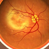 |
Description: Video illustration of changes in the eye associated with AMD. Age-related Macular Degeneration (AMD) is the leading cause of visual impairment and blindness in older Americans. It affects the retina, the light sensitive layer of the eye. As yellowish deposits form under the retina, they can result in distortion and gradual blurring of vision. This is called "dry AMD." The second type, called "wet AMD" can lead to bleeding and more rapid vision loss. The most common form is the dry type, but as more and larger deposits develop under the retina, the risk of developing the wet type increases. Audio Description: (Video only; no audio.) The animation begins with a close-up of the face of an elderly woman. The face fades as the camera begins to zoom in on her right eye. The zoom continues and as the eye fills the screen, the front half disappears to reveal the light sensitive retinal layer at the back of the eye. Small yellowish deposits known as drusen are seen forming under the retina blurring the sharp central area of vision or macula. Individual drusen, coalesce forming larger areas of damage. Blood vessels growing up from below the retina leak blood under the retina. Pressure from these pockets of blood, damage the light sensing cells, destroying the ability to see straight ahead. Credit: National Eye Institute, National Institutes of Health View Animation (Quicktime) Ref#: VA03 |
|
 |
Description: Video illustration of changes in the eye associated with AMD combined with a vision simulation. Age-related Macular Degeneration (AMD) is the leading cause of visual impairment and blindness in older Americans. It affects the retina, the light sensitive layer of the eye. As yellowish deposits form under the retina, they can result in distortion and gradual blurring of vision. This is called "dry AMD." The second type, called "wet AMD" can lead to bleeding and more rapid vision loss. The most common form is the dry type, but as more and larger deposits develop under the retina, the risk of developing the wet type increases. Audio Description: (Video only; no audio.) The animation begins with a close-up of the face of an elderly woman. The face fades as the camera begins to zoom in on her right eye. The zoom continues and as the eye fills the screen, the front half disappears to reveal the light sensitive retinal layer at the back of the eye. Small yellowish deposits known as drusen are seen forming under the retina blurring the sharp central area of vision or macula. Individual drusen, coalesce forming larger areas of damage. Blood vessels growing up from below the retina leak blood under the retina. Pressure from these pockets of blood, damage the light sensing cells, destroying the ability to see straight ahead. The progress of AMD is repeated with the development of drusen and later, pockets of blood under the retina, as shown in the inset in the upper right. The main image is a simulation of what the individual sees as the disease progresses. AMD disease progress and gradual vision loss are presented concurrently in the last part of the animation. Credit: National Eye Institute, National Institutes of Health View Animation (Quicktime) Ref#: VA04 |
|
 |
Description: A child with amblyopia is sending unequal signals from each eye to the brain. As shown in this animation, neuro-electrical signals travel along pathways from the eye to the brain. The unaffected right eye is sending strong signals. The eye with amblyopia, the left eye, is sending fewer neuro-electrical signals. If untreated, the pathways through which these signals travel may weaken and not develop properly, damaging the child’s vision.
Placing a patch over the unaffected eye for several weeks will stimulate and strengthen the signals from the eye with amblyopia leading to more normal nerve function in the brain, which improves vision in that eye. Credit: National Eye Institute, National Institutes of Health View Animation (14 second MPEG) View Animation (14 second Quicktime) View Animation (30 second Quicktime) Ref#: VA02 |
|
 |
Description: Video illustration of retinopathy of prematurity ROP: ROP develops when normal blood vessel growth stops in the retina, nerve tissue that lines the back of the eye. In the worst situation, the normal blood vessels are replaced by abnormal vessels, which then may spread throughout the retina and into the center of the eye (vitreous). The scarring and bleeding may lead to retinal detachment, resulting in severe vision loss. Audio Description: (Video only; no audio.) A close up of an infant's face. The camera begins to zoom in on the child's left eye. The zoom continues until the eye fills the screen at which point the image morphs into an animation of the child's eye. The animation then rotates to reveal a side cross-section of the child's eye. The animation continues illustrating abnormal blood vessel development leading to scarring, bleeding and possible retinal detachment. Credit: National Eye Institute, National Institutes of Health View Animation (MPEG) Ref#: VA01 |
|
TOP

