NIBIB Calls for Substantial Reduction in Radiation from Routine CT Scans
How much radiation in medical tests is too much? It’s a complicated question with no easy answers. However, the one thing most physicians and researchers agree on is that the less radiation involved in diagnosing and treating patients, the better. The National Institute of Biomedical Imaging and Bioengineering (NIBIB) is taking a central role on an initiative to develop new methods and technologies for reducing the radiation dose from routine CT scans while maintaining the high quality of the images produced.
As a part of this initiative, NIBIB has put out a call to researchers for groundbreaking ideas that will help to radically decrease the amount of radiation used in CT scans. This new funding opportunity is intended to inspire innovation and creativity and allow researchers to try new approaches that might not be funded otherwise.
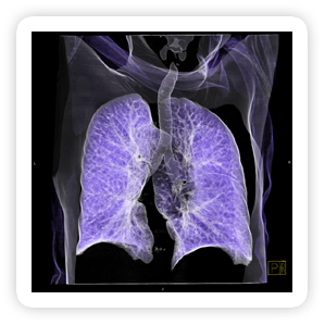
NIBIB began to concentrate its focus on CT radiation in February of 2011 as the lead sponsor of an experts’ summit on how to reduce radiation exposures from CT studies while still maintaining the
 |
Fig 1. Volume CT of Pulmonary System
Click on image to enlarge |
highly detailed image information that physicians need from these critical studies.Participants included both technological and clinical specialists from academia, industry, and government regulatory and funding agencies. The driving goal of the meeting, according to NIBIB Director Roderic I. Pettigrew, Ph.D., M.D., was to identify what would be required to reduce patient exposure from a routine CT examination to less than 1 millisievert (1 mSv). A mSv is a measure of how much ionizing radiation is absorbed by a specific volume of tissue from an exposure to X-rays. Typical doses from CT studies range from 1 to 12 mSv, with an average dose of 6.5 mSv, (which is equivalent to the dose received from more than 300 chest X-rays). Experts at the summit discussed possible strategies for how current, soon-to-be-available, and future technologies could be expected to collectively achieve CT examination radiation doses of less than 1 mSv.
Attendees also discussed possible new funding opportunities, identifying areas in which technology is already rapidly progressing, such as the development of new x-ray sources and beam
Not all medical imaging studies involve radiation, but computed tomography, x-ray, mammography, and angiography all use radiation. Computed tomography (CT), is a crucial medical technology accounting for only 17% of imaging studies, but nearly 49% of medical radiation exposures
1.
configuration, innovation in detectors, automatic exposure control, and new methods for computer processing and reconstruction of images from the detected X-ray data. Another key factor identified was reducing background noise. CT “noise,” is a term that refers to the degradation of the X-ray image resulting from insufficient X-ray data reaching the detectors. This type of image degradation can hamper the correct interpretation of a CT study. If successful, these technological improvements could help decrease the amount of radiation required to produce a good CT image.
In addition to technological improvements, there are other steps that can be taken now to decrease radiation from CT studies. Summit participants were unanimous in their enthusiasm about
 |
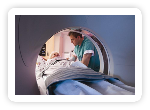
Fig 2. CT Scanner
Click on image to enlarge |
physician training as a feasible means to immediately reduce radiation exposure. “Dose reduction does start with dose prevention,” said Dr. Mannudeep Kalra, MD, an assistant radiologist at Massachusetts General Hospital. While a CT scan might at first be deemed clinically necessary, physician training on imaging advances may challenge clinicians to consider alternative scans. Physicians should choose the best method for the level of imaging sensitivity needed for a particular medical question. Depending on the purpose of the scan, the dose can vary dramatically, so doctors need to be aware of how much radiation their patients will receive as a result of a CT exam. Expanded training could help physicians who order CT studies better understand when a non-ionizing radiation based scan, such as magnetic resonance imaging (MRI) or ultrasound, could be used instead.
“When a CT scan is clinically warranted, the benefits by far outweigh any possible individual radiation risk,” said Hedvig Hricak, MD, PhD, Chair of the Department of Radiology at Memorial Sloan-Kettering Cancer Center. Whereas radiation exposure is regulated in the workplace, medical radiation has almost no oversight, with the exception of mammography. When the Mammography Quality Control Standards Act was passed in 1992, Dr. Hricak noted that it was initially annoying to doctors. However, the regulation did drastically decrease overuse of the test and, Dr. Hricak pointed out, “in the end everybody is better off.”
1Mettler FA Jr., Bhargavan M, Faulkner K, et al. Radiologic and nuclear medicine studies in the United States and worldwide: frequency, radiation dose, and comparison with other radiation sources—1950–2007. Radiology 2009;253(2):520–531.
 More Highlight on Health articles
Last Updated On 09/13/2012