Electron microscopy (EM) of stained cell sections is used to definitively localize complex molecular assemblies within cells (e.g., chromatin, organelles and membranes). This technique also can be used to determine protein structures. NIGMS supports grants that apply EM as a structural and biophysical method to study any basic cellular process, as well as to develop molecular tags that can be genetically targeted and eventually used for in vivo imaging. Here are a few examples of projects in this area.
Localization of Organelles by EM
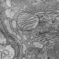
 Cells were fixed, stained, dehydrated and embedded in plastic in order to be cut thinly for imaging in the transmission electron microscope. This image of a thin section of a single cell shows distinct cellular compartments and structures within them. Credit: Tina Carvalho, University of Hawaii.
Formation of the “Bullet” Tip of VSV Virion
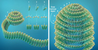
 This is a cartoon illustration based on cryoEM density data of a plausible process by which the vesicular stomatitis virus nucleocapsid ribbon generates the virion head, starting with its bullet tip. The curling of the nucleocapsid ribbon generates a decamer-like turn at the beginning, similar to the crystal structure. When assembly nearly completes this turn, continuation of vRNA requires that the ribbon form a larger turn below it. As the spiral enlarges and progresses to the helical trunk, the tilt of individual subunits decreases. When it reaches the 7th turn, the nucleocapsid ribbon becomes helical, with each new turn of the nucleocapsid fitting under the preceding one. Credit: Z. Hong Zhou, University of California, Los Angeles. Article abstract