

Visionary
Studying the Lens of the Eye with MRI
Interview with Susan Strenk, Ph.D.
By Allyson T. Collins, M.S.
NEI Science Writer/Editor

Susan Strenk, Ph.D.
Vice President
MRI Research, Inc.
Her undergraduate degree in electrical engineering and Ph.D. in biomedical engineering may seem a bit impersonal, but Susan Strenk spends most of her time on intimate encounters--staring into people's eyes.
She does it with the help of MRI, or magnetic resonance imaging, which uses radio-frequency pulses to provide a detailed look inside the organ. Strenk and her husband Lawrence, an MRI physicist, founded MRI Research, Inc. and have spent the past six years studying the eye's natural lens and artificial lenses implanted during cataract surgery with the help of funding from the National Eye Institute's Small Business Innovation Research (SBIR) program.
In a recent interview, Strenk talks about turning her fascination with eyes into a business built around them.
When did you become interested in eye research?
I've always had a strong interest in eyes. In fact, as a junior high student, I asked to take home the goat eye that we dissected. I finally had an opportunity to study eyes in a scientific way when I was in graduate school. My research involved looking at how the eye focuses using MRI. I was responsible for building small radio-frequency receiver coils to give us a higher image resolution in a small area.
How does MRI work?
"SBIR grants make our research possible," says Strenk. "These grants are virtually the only source of seed money for innovative, high-tech businesses."The MRI machine contains a very strong magnet. When people go into the machine, the hydrogen atoms in their body line up with the magnetic field. The machine applies a radio-frequency pulse, which is not harmful like the radiation used in X-rays and CT scans. That pulse knocks the hydrogen atoms out of alignment. After the pulse, the atoms realign and release a signal that's picked up by a receiver coil, which is like an antenna. A computer translates these signals into images.
Does MRI give you a better look at the eye than other imaging methods do?
MRI is unique in letting us look behind the iris, which is the colored part of your eye. We can identify different eye structures very well by the different shades of gray in the image. For example, the iris appears brighter than the surrounding fluid. We can also get images from any angle, so we can see the entire lens and the ciliary muscle, which contracts and relaxes when we focus on near and far objects--a process called accommodation. Before MRI, no one had actually seen the ciliary muscle in the normal, living human eye because it is blocked by the iris, except in a few rare cases where the iris was missing.
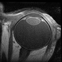
MRI of a 63-year-old man's healthy eye (courtesy of MRI Research, Inc.)
Why did you decide to start a company based on this technology?
The most common surgical procedure in the world is cataract surgery, where surgeons remove the natural lens of the eye and replace it with a synthetic intra-ocular lens (IOL). We founded MRI Research, Inc. in 2003 to use MRI to provide feedback for companies on how IOLs respond when the eye tries to accommodate. We applied for our first SBIR grant from the National Eye Institute that year, so we could build a database of MRI measurements from cataract patients and donor eyes.
Why did you apply for SBIR funding?
These grants are virtually the only source of seed money for innovative, high-tech businesses. Banks will not give loans to start small, high-risk businesses, and the venture capital industry will not invest at such an early stage. SBIR grants make our research possible.
What types of studies have you done with this support?
Our initial Phase I study involved imaging 10 people with IOL implants while they looked at close and far away objects. We took measurements of many structures in the eye to document changes in their shape and location when the eye accommodates. Our phase II study is tracking more patients during the year after surgery. One of the long-term effects we're focusing on is Soemmering's ring, a tissue that can develop around part of the IOL and change its location, decreasing vision quality. We are also looking at donor eyes, which help us get much more detailed images than we could from cataract patients because we can image for longer periods of time. By combining measurements from patient and donor eyes, we get a much more complete picture of what's happening.
What do you hope to accomplish through your research?
Our main goal is to help better IOLs reach the clinic, such as accommodating lenses that restore the ability of people to focus at different distances when they age and develop presbyopia. We're also helping medical professionals and device manufacturers learn more about how cataract surgery affects other parts of the eye. Ophthalmologists recently found that the procedure lowers eye pressure in patients who had high pressure before surgery. Our images have helped show that, with age, natural lenses grow and push the iris forward, possibly pinching off a small canal that helps maintain normal eye pressure. The IOL may actually return the eyes of these patients to a relatively youthful state.
References:
Strenk SA, Strenk LM, Guo S. Magnetic resonance imaging of the anteroposterior position and thickness of the aging, accommodating, phakic and pseudophakic ciliary muscle. Journal of Cataract Refractive Surgery, February 2010.
Strenk SA, Strenk LM, Koretz JF. The mechanism of presbyopia. Progress in Retina and Eye Research 2005; 24: 379-393.

