

Insight
Big Ideas from Small Businesses
Vision-related research by innovative thinkers
By Allyson T. Collins, M.S.
NEI Science Writer/Editor
A printer that produces high-resolution graphics, which can be "viewed" through the touch of a visually impaired person; a high-resolution imaging system for doctors to examine the eyes of premature infants; and eyeglass lenses that expand the sight of people who have lost half of their vision.
"These researchers are taking great ideas and turning them into real products that people can use," Wujek says.These are just a few of the nearly 90 projects currently funded at $14 million by the National Eye Institute, through a special grants program called Small Business Innovation Research (SBIR). The NEI's Jerome Wujek, Ph.D., oversees these grants and tracks the progress of the start-up companies behind them.
"The SBIR program covers the whole waterfront, from vision assistive devices to medications to ophthalmic instruments and everything in between," Wujek says. The focus of these efforts is on product commercialization.
To achieve this, the SBIR program is structured in three phases. During Phase I, businesses are given funding for developing a prototype and determining whether a proposed product will work. Phase II funding is for the heart of the project: turning the prototype into a product and proving that it works effectively and safely. Phase III is not funded by the NEI, but is the process by which the project should be advanced enough to allow commercialization, which could include clinical testing and Food and Drug Administration (FDA) approval.
"Through SBIR grants, small businesses can move the field forward with out-of-the-box thinking," Wujek says. "These companies are taking great ideas and turning them into real products that people can use."
Gaining access to graphics
"One day, I went completely blind," says John Gardner, Ph.D.
It was in 1988, after Gardner had surgery for glaucoma--the rising eye pressure that had steadily decreased his vision. There were surgical complications, and when the bandages were removed, his world was dark.
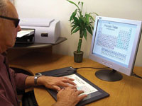
John Gardner, Ph.D., president of ViewPlus Technologies, Inc., uses the IVEO Viewer and touchpad to explore a periodic table of the elements (courtesy of ViewPlus Technologies, Inc.)
The 48-year-old underwent additional surgeries, but his vision didn't return. "I never quit working, though," he says. "I had too much to do."
Gardner was a physics professor at Oregon State University. When he returned to his office, he used a computer screen-reader and a textbook scanner, but a crucial part of his career couldn't be translated.
"I had access to words, but I had no way to do math for my research," Gardner explains. "And an even worse problem was that I couldn't see charts or graphs." When his wife handed him a tactile picture that he should have been able to touch and describe, he thought it was two pizza pans and a lot of wires. She told him it was a bicycle.
Gardner was confident that he and his colleagues could design a technology for embossing high-quality, high-resolution graphics on paper at 20 dots per inch--the resolution that a human finger needs to "read."
One of his students achieved that goal in 1996, creating TIGER, a tactile graphics embosser technology that produces high-resolution, three-dimensional graphics. They started ViewPlus Technologies, Inc. shortly thereafter, and applied for an NEI SBIR grant in 2002 to develop a combination hardware-software program that would read onscreen graphics and translate them for visually impaired people.
Through SBIR grants, they built the IVEO Viewer, a program that works with a touchpad, so users can feel and hear information about computer graphics. They also created a printer that produces tactile copies in both ink and Braille--allowing better communication between blind and sighted people.
Gardner's group is encouraging universities and libraries to purchase the printers, so visually impaired visitors can have access to graphics, such as maps in history books.
"It is now possible for anybody who makes graphical information to provide it in a form that is automatically accessible to everybody," Gardner says.
Imaging the eyes of infants
In the early 2000s, Eric Buckland quit his job in the telecommunications industry to apply his Ph.D. in optics to life sciences.
"In the community of physicians, researchers, and patients, there's a degree of passion that I hadn't seen in the telecommunications space," Buckland explains. He connected with Joseph Izatt, Ph.D., who was working at Duke University on optical coherence tomography (OCT).
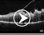
Similar to the way that ultrasound uses sound waves to generate images of body organs, OCT uses light waves to capture high-resolution images of living tissues. An OCT system for the eyes requires people to place their head on a chin rest and hold still for several seconds as the images are obtained. The process is painless, and the system doesn't touch the eye.
At the time, Buckland says, many routine OCT systems existed, but there were no high-performance systems for OCT-based eye research. "The research community was asking for these tools," he explains.
Buckland and Izatt co-founded Bioptigen, Inc. in 2004. They used NEI SBIR funding to develop a versatile OCT system that includes interchangeable probes for scanning different sizes of eyes and locations within the eye--from the cornea in the front to the retina in the back. The system received FDA clearance in 2006.
Researchers initially used the system's hand-held probe to image the eyes of animals. Then, they realized that they could also use it during eye surgeries and for diagnosing infants and toddlers with eye conditions, such as retinopathy of prematurity.
"There is only one other imaging instrument on the market dedicated to pediatric eye care, and no other OCT system," Buckland says.
Bioptigen's OCT system is now being used in research laboratories like the Medical College of Wisconsin, in clinical trials such as an OCT study managed by Duke University as part of the NEI-supported Age-Related Eye Disease Study 2, and in pediatric clinics like Childrens Hospital Los Angeles, among other locations.
"What the SBIR grants allow us to do is address some of the underserved areas of ophthalmology, such as pediatrics," Buckland explains. "Knowing that our system can play a role in saving the eyesight of young children is very satisfying."
Designing lenses
As a child, Karen Keeney, M.B.A., lived with her grandfather, who was legally blind.
"I became aware of visual problems on a first-hand basis," she says.
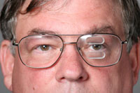
A man wears glasses with the Peli lens (courtesy of Chadwick Optical, Inc.)
As an adult, Keeney worked at American Optical, a lens company where she met her "optical mentor," John Chadwick. He showed her how to manage inventories and market products, leading her to purchase an optical laboratory in 1980.
Chadwick Optical, Inc. made traditional eyeglass lenses until the late 1990s, when corporations began to dominate the field. "With all the strength and money these big labs had, small labs like mine were doomed," she explains.
So she picked a niche market--low vision--and started designing prototype lenses. She sent drawings to researchers, including NEI-grantee Eli Peli, M.Sc., O.D., at Schepens Eye Research Institute in Boston.
"He called me and told me, 'You have to design lenses around a problem you're trying to solve, not design lenses and fit them to a problem,'" Keeney remembers.
But he appreciated her initiative, so the two met in 2002 and sketched out the "Peli Lens." It was designed for people with hemianopia who have lost half their vision--as if someone put post-it notes over the left or right side of both eyes, Keeney explains. The condition can be caused by a stroke, trauma, or brain damage.
Peli lenses use high-powered prisms to draw some lost vision into view. They have prisms above and below a person's line of sight, so they don't interfere with normal vision, and are generally used only on the side where a person is blind, Keeney says.
Users see blurred images in the prisms. If a man wearing prisms on the left side walks toward a door, the left doorjamb will appear blurry above and below his central vision. He can then turn to see the doorjamb clearly.
"These lenses create an artificial peripheral vision," Keeney says. "The blur actually helps people detect what's real and what's shifted."
Through NEI SBIR funding, the lenses were developed and tested in a clinical trial, which found that 74 percent of patients continued to wear them after six weeks. Keeney and Peli are now testing larger prisms in another clinical trial.
"There is no training involved," Keeney says about the lenses. "People generally get the hang of it right away."

