

Insight
Subtle Saccades
Researchers learn about the brain through quick eye movements
By Allyson T. Collins, M.S.
NEI Science Writer/Editor
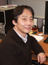
Okihide Hikosaka, M.D., Ph.D.
Chief, Neuronal Networks Section
NEI Laboratory of Sensorimotor Research
Okihide Hikosaka, M.D., Ph.D., can't quite put his finger on why he became interested in neuroscience. When asked, he sits for a moment, smiles, and shrugs, as if it's natural for him to love learning about how our brain moves our eyes and how, whether we realize it or not, our emotions have some control over this process.
His lack of an explanation doesn't phase him. He's faced with unanswered questions every day. As chief of the Neuronal Networks Section in the NEI Laboratory of Sensorimotor Research, he manages a six-person lab, which investigates the complex coordination of nerve cells that results in eye movements.
After discussing his work, however, he revises his prior silence. "The reason why I was attracted to neuroscience is probably its seeming ability to produce something, like creativity, from nothing," he says. "I still believe that there must be some nerve cells in the brain that cause creativity, but we haven't found them yet. I'm still puzzled by that. But in the meantime, I have been studying eye reflexes and movements."
Bursting with energy
Hikosaka's first experience with eye research was during his graduate work at the University of Tokyo, where he studied the relationship between equilibrium and eye movements.
When many species rotate their heads, their eyes keep looking in the same direction. He describes this by pretending that a frog is sitting on a piece of paper in front of him. If he turns the paper, he explains, the frog's head turns in the opposite direction so that the frog will keep looking straight at him.
"Vision is the first thing that many of us use to search for something and analyze the world," Hikosaka says."The purpose of this reflex is to stabilize the image on the retina, otherwise the visual system can't capture the image," Hikosaka says. Our eyes are like a camera that has a slow shutter speed. If the scene changes quickly, the image will be blurry. Because of this, people who have problems maintaining balance also have visual problems, he says.
That reflex was the starting point of his research, but he was eager to look more at what happens when a reflex causes the eye to move. Back to the frog, he explains that when the paper rotates past a certain point, near 90 degrees, the frog's head will adjust to look at the wall, instead of at him. This quickly resets the frog's visual image.
Researchers understood that these movements, known as quick-phase reflexes, originated in the brainstem area, but Hikosaka was the first to identify a group of nerve cells called burst neurons that make this movement possible.
"Those neurons emit a burst of electrical energy that is sent to motor neurons, which control eye muscles," he explains. "The motor neurons receive the burst signal and create a twitch in certain muscles and a relaxation in others to allow your eye to rotate quickly."
Searching through saccades
Though Hikosaka remained captivated by burst neurons, he wanted to move beyond innate reflexes to know what happens when animals control their eye motion, a process known as saccadic eye movement.
"Quick-phase movement is a reflex, but at some point during evolution, neurons in another area of the brain probably gained connections to burst neurons," he says. He explains that those other neurons receive information about the sights and sounds around us. The burst neurons use that visual or auditory information to allow an animal to make voluntary saccadic eye movements.
We make tiny, imperceptible saccades when we look at images and even when we walk into a room. "You might not be aware of how you move your eyes, but you're probably making saccades two to three times per second," Hikosaka says. "Vision is the first thing that many of us use to search for something and analyze the world."
After finishing his Ph.D. in Tokyo, Hikosaka came to NEI in 1979 to do research solely involving these voluntary saccades. Here, he discovered neurons in another area of the brain, called the basal ganglia, which keep burst neurons from being continually activated, to prevent constant eye movement.
He returned to Tokyo to continue working on "closing the loop" of brain activity related to saccadic eye movement. Twenty years later, a permanent position at NEI opened and he jumped at the chance to return.
"Back in Japan, as a professor, I had little time working in the lab because of teaching and administrative duties," he says. "After coming back here, I can spend much more time experimenting, which is crucial."
Tackling a variety of topics

Hikosaka and Hong showed that when a subject sees a reward or punishment, a brain circuit is activated that energizes or slows eye movements.
Hikosaka's research at NEI spans a wide range of areas. "My long-term goal is to understand the neural mechanisms of voluntary behavior--how you initiate your movements based on your will," Hikosaka says.
Among his many projects, he researches the role of a brain structure called the lateral habenula, which may be involved in the origin of depression and schizophrenia, and collaborates with Japanese scientists to study eye movements of patients with Tourette syndrome.
Three years ago, Simon Hong, Ph.D., asked to join Hikosaka's lab to study how emotions, particularly disappointment, affect eye movement. Hikosaka's team had already found that neurons in the lateral habenula can stop dopamine--a chemical neurotransmitter in the brain--from being released.
"Dopamine is the substance that energizes the body to do a particular task," Hong says. "When dopamine is not available, the energizer of the body is shut off, making us feel disappointed and sluggish."
Hikosaka and Hong identified the first step in the brain circuit that causes the lateral habenula to become activated, preventing dopamine production. They also noticed that the lack of dopamine causes the eyes to move more slowly, decreasing the speed of saccades.
"Our animal subjects move their eyes slower when they are disappointed, as opposed to the energized, ballistic movements they make when they are motivated," Hong says. "We found that the emotional circuit interacts with the motor circuit to make it move faster or slower."
Hong says that understanding how brain circuits involving emotions can control our behaviors could help researchers identify treatments for neurological disorders such as Alzheimer's and Parkinson's diseases. But for now, Hikosaka and his team keep their eyes focused on the laboratory bench.
"If you aim at clinical applications at the beginning, your chance of finding something new may be limited," he says. "If you maintain all different types of basic research, you will have more opportunities to find something really unexpected that could be very useful in clinical therapy."
References:
Bromberg-Martin ES, Hikosaka O (2009) Midbrain dopamine neurons signal preference for advance information about upcoming rewards. Neuron 63:119-126.
Hong S, Hikosaka O (2008) The globus pallidus sends reward-related signals to the lateral habenula. Neuron 60:720-729.
Matsumoto M, Hikosaka O (2009) Representation of negative motivational value in the primate lateral habenula. Nat Neurosci 12:77-84.
Matsumoto M, Hikosaka O (2009) Two types of dopamine neuron distinctly convey positive and negative motivational signals. Nature 459:837-841.
Matsumoto M, Hikosaka O (2007) Lateral habenula as a source of negative reward signals in dopamine neurons. Nature 447:1111-1115.

