
June, 2009
Insight
The Mystery of Healthy Eyes
Understanding normal vision before treating disease
By Genevive Bjorn, M.S.
NEI Freelance Science Writer
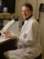
Peggy Zelenka, Ph.D.
Chief, Laboratory of Molecular and Developmental Biology
Head, Section on Cellular Differentiation
National Eye Institute
More than 3 million Americans age 40 and older experience low vision or blindness, but doctors still lack effective treatments for many sight-stealing conditions. Finding therapies for treating diseased eyes often depends on first understanding how normal eyes work, a familiar concept for the chiefs of two National Eye Institute (NEI) laboratory sections.
In the Laboratory of Molecular and Developmental Biology, Stanislav Tomarev, Ph.D., and Peggy Zelenka, Ph.D., study how healthy eye cells grow, develop and communicate with neighboring cells.
"We are trying to understand normal physiology first, then apply that to disease," Zelenka explains.
Mixed Signals
Glaucoma is a leading cause of blindness in the United States. In many cases, this condition is associated with high pressure inside the eye that damages the optic nerve, interrupting transmission of information from the eye to the brain.
Sometimes glaucoma is caused by problems with a protein called myocilin, which is part of a large group of related proteins. Myocilin is involved in fluid drainage in the front of the eye, an important process for maintaining healthy eye pressure.
In 2008, Tomarev's group looked at a related protein called olfactomedin 1 in tropical, freshwater zebrafish and found that this protein regulates optic nerve cell development and growth in the eye.
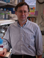
Stanislav Tomarev, Ph.D.
Chief, Molecular Mechanisms of Glaucoma Section
Laboratory of Molecular and Developmental Biology
National Eye Institute
Therefore, they reasoned that an abnormal olfactomedin 1 protein could result in damage to the optic nerve, which could increase the risk of developing glaucoma.
"With this information, we're trying to figure out ways to prevent the disease process that is initiated by a damaged protein," Tomarev explains. "We're also trying to identify treatments that could eliminate this vision-threatening effect."
Refreshing Vision
Zelenka and her group study the lens and cornea, where they trace signals that trigger cell growth and activity. Cells in the center of the lens never replenish and must remain clear throughout a person's lifetime--a daunting physiological challenge. When transparent lens cells become cloudy, cataracts can develop.
Fortunately, the outer part of the lens constantly receives a fresh supply of transparent cells that help support the aging cells within. These new cells are generated from a small population of cells at the edge of the lens through a process known as differentiation.
Researchers in Zelenka's lab have recently shown that signals from a protein called Notch are required for this complex process of lens cell differentiation to proceed normally. This contradicts what previous researchers had found--that signals from Notch actually blocked lens cell differentiation.
"With this discovery, we realized that Notch works in two different ways," Zelenka says. "Notch prevents cell differentiation in the front of the lens. But when lens cells migrate to the back part of the lens--the normal site of differentiation--they receive signals that actually change the Notch signal to one that promotes differentiation."
Another project in Zelenka's lab focuses on an enzyme known as Cdk5, which is one link in a chain of events that helps lens and cornea cells hold tightly to their basement membrane. This membrane is formed from the cells' natural secretions.
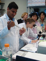
Scientists at work in the Section on Cellular Differentiation in the NEI Laboratory of Molecular and Developmental Biology.
Adherence to this membrane is crucial for cell survival; cells will die if they detach from their basement membrane. However, this property can also be detrimental in certain situations, such as when cells need to move quickly to heal a damaged cornea.
When Zelenka's team blocked the Cdk5 enzyme in corneal cells, cell migration speeded up. "Based on this observation, we think that cells might have natural ways of reducing Cdk5 when they need to switch locations," Zelenka explains.
Finding Treatments
While they continue to uncover new information about healthy eyes, scientists in both Tomarev's and Zelenka's laboratory sections are applying these discoveries to develop treatments for eye diseases.
Tomarev's team, in collaboration with scientists at Cambridge University in the United Kingdom, is exploring the use of stem cells. They began by looking at retinal tissue in cultures, which allowed them to identify the best conditions for the stem cells to integrate into existing retinal tissue. This method is also simpler and faster than animal studies, Tomarev says.
They cultured stem cells with normal cells isolated from the retina and with cells that had been altered to simulate changes observed in glaucoma. They discovered that the presence of stem cells actually delayed cellular changes that lead to glaucoma. More recently, they moved to animal studies, where they confirmed these results.
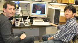
Graduate student Thomas Johnson (L) and Naoki Nakaya, Ph.D., analyze a section of rat retina through an epifluorescent microscope in Tomarev's laboratory. The microscope allows them to study retinal and brain cells that have been labeled with fluorescent markers.
"We tried just one type of stem cells," Tomarev says. "There are many other types of stem cells that we could use. In the future, we will test the effectiveness of other stem cell types as well, including genetically-modified stem cells."
In Zelenka's lab, researchers recently made eye drops containing a substance that temporarily shuts down the effectiveness of Cdk5. When they tested the drops on mice that had damaged corneas, they found that wounds closed more rapidly because the cells migrated faster than when Cdk5 kept them in their normal location. Not only did the wounds heal more quickly, but they also healed without a common side effect: inflammation.
"We need more studies to determine whether these drops could be effective in humans," Zelenka says. "Still, we hope that someday, similar drops could be used to treat people who have diabetes because they tend to heal more slowly than others."
References:
Johnson TV, Martin KR (2008). Development and characterization of an adult retinal explant organotypic tissue culture system as an in vitro intraocular stem cell transplantation model. Invest Ophthalmol Vis Sci, 49, 3503-12.
Kwon HS, Lee HS, Ji Y, Rubin JS, Tomarev SI (2009). Myocilin is a modulator of Wnt signaling. Mol Cell Biol, 29, 2139-54.
Ledee DR, Tripathi BK, Zelenka PS (2007). The CDK5 activator, p39, binds specifically to myosin essential light chain. Biochem Biophys Res Commun, 354, 1034-9.
Nakaya N, Lee HS, Takada Y, Tzchori I, Tomarev SI (2008). Zebrafish olfactomedin 1 regulates retinal axon elongation in vivo and is a modulator of Wnt signaling pathway. J Neurosci, 28, 7900-10.
Saravanamuthu, SS, Gao, CY, and Zelenka, P (2009). Notch signaling is required for lateral induction of Jagged1 during FGF-induced lens differentiation. Develop. Biol. (in press).
Tripathi BK, Stepp MA, Gao CY, Zelenka PS (2008). The Cdk5 inhibitor olomoucine promotes corneal debridement wound closure in vivo. Mol Vi, 14, 542-9.

