
September 2009
Snapshot
Damaged Rods and Cones
Retinitis pigmentosa impacts light-sensitive cells
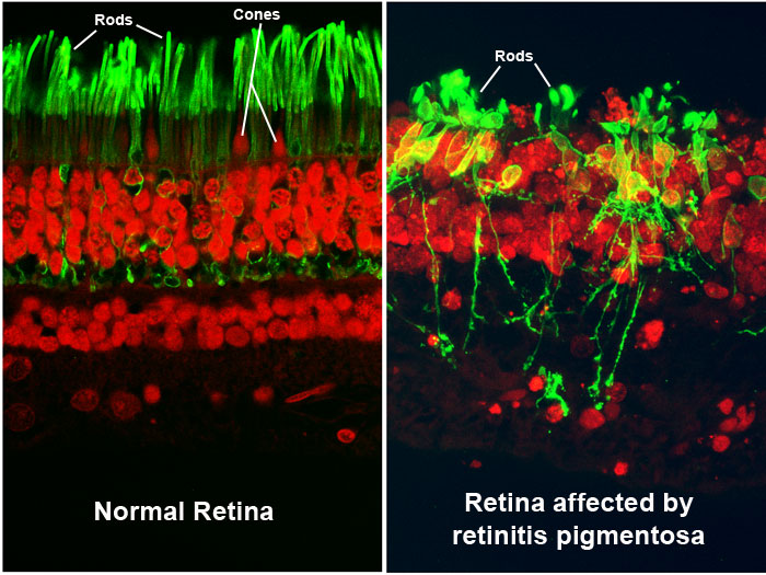
These images show a section of retina tissue from the back of the eye, as seen through a high-resolution microscope. The left image illustrates a normal retina, while the right image shows a retina affected by a condition called retinitis pigmentosa (RP).
RP is a genetic eye condition. People who have RP first experience gradual vision loss in their side vision and night vision, and later in their central vision. They may eventually become blind. Currently, there is no effective treatment to prevent vision loss from RP.
RP damages the retina's light-sensitive photoreceptor cells, which connect with other nerve cells to transmit visual information to the brain. Rod photoreceptors that control night vision are impacted most often, but cone photoreceptors involved in color vision can also be affected by RP.
In both images, the rods are shown in green. In the normal retina on the left, the rods and cones are organized in an orderly fashion, with the circular red cones in between the rods. In the RP retina, this organization is disrupted as the retina begins to deteriorate. The damaged photoreceptors invade other areas of the retina, causing them to lose their connections with other nerve cells. This invasion process as well as the death of photoreceptors can cause the vision loss associated with RP.
Image courtesy of Robert N. Fariss, Ph.D., chief of the NEI Biological Imaging Core, and Ann H. Milam, Ph.D., former professor in the Department of Ophthalmology at the University of Washington.

