
October 2009
Snapshot
The Eye's Relay Race
Visual information transmitted between neurons
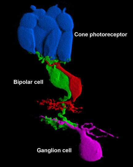
This image shows three major types of nerve cells in a slice of retina tissue from the back of the eye. The nerve cells, or neurons, work together to process visual information and transmit it to the brain.
Researchers injected different fluorescent dyes into each of the neurons with a tiny needle-like instrument called a micropipette. Then, they used an imaging tool known as a confocal microscope to detect the cells' fluorescence and produce a high-resolution image.
The blue-colored neurons are cone photoreceptors involved in color vision and vision in brightly lit environments. Cones convert light energy into electrical signals. These electrical signals are communicated to the green- and red-colored bipolar cells with the help of a chemical known as a neurotransmitter.
Bipolar cells are like the middleman in a relay race--they receive input from photoreceptors at one end, and hand off the signals to ganglion cells at the other end. The magenta-colored ganglion cells have long nerve fibers called axons that come together to form the eye's optic nerve, which carries visual information to the brain for further processing. The brain ultimately generates the image that is seen.
Image of ground squirrel retinal tissue courtesy of Wei Li, Ph.D., chief of the NEI Unit on Retinal Neurophysiology.

