 NCI NewsCenter
 NCI Budget Data
 Visuals Online
 NCI Fact Sheets
 Understanding Cancer Series
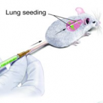
Tumor cells were delivered to mice by tail-vein injection.
Metastasis of a tumor from its primary site to other parts of the body continues to be the main reason why people die from cancer. In a new technical advance, NCI scientists have successfully designed, implemented, and validated a model that allows scientists to explore—in real-time—the progression of cancer as it metastasizes in the mouse lung. Understanding the mechanisms of how metastasis works is an important step in stopping its progression.
Assay Follows Metastatic Progression of Tumor Cells to a Secondary Site
An informal collaboration of investigators at the NCI has led to the formation of the Center for Cancer Research Metastasis Working Group. The group discussed the challenge of doing metastasis research since most of the process occurs in a black box, i.e. it is done in animals, such as mice, making it challenging for scientists to follow the metastasis that occurs within the mice once they are inoculated with tumor cells.
The group theorized that introducing tumor cells into animals just prior to performing a lung culture might allow them to study the metastatic progression as it was happening. With this aim in mind, they developed an ex vivo (PuMA) in which cancer cells marked with a fluorescent molecule can be viewed during metastatic progression—from a single tumor cell to a secondary site in the lung. The scientists hope this technical advance will help clarify the steps of metastasis that scientists have primarily been able to theorize about, or model poorly, in other in vitro systems.
The scientists acknowledge that there are other approaches to studying metastasis in animals, in real-time, but explain that those approaches require microscopic tools that are not widely available and not amenable to the study of the lung. The PuMA allows the serial assessment of metastatic progression without such instruments and provides a new window into the steps involved in metastatic progression.
Technical Advance—Modeling Metastasis Biology and Therapy in a Real-Time Mouse Lung
In this experiment, the scientists compared a series of high and low-potential metastatic cells both in mice and in the PuMA. Malignant tumors are composed of cells with different characteristics and only certain cells have metastatic potential; the establishment of cell lines with high or low metastatic potential is necessary to investigate the molecular mechanisms of the metastatic process. The results of the study were published in The Journal of Clinical Investigation, in August 2010.
Tumor cells were labeled with a fluorescent marker and then introduced into the mouse’s circulation by tail vein injection. Subsequently, the lungs were excised, filled with agarose—a type of complex carbohydrate—to provide structural support, sliced, and placed in culture. This allowed the researchers to follow the growth of the tumor cells over time, and to view growth from single cells to proliferating cell colonies. In each case the cells that were highly metastatic in mice were also highly metastatic in the PuMA. The same was true for low metastatic cells.
With this validation in place, the team showed that the assay also provided an opportunity to evaluate new cancer drugs for their specific effects on metastatic progression. This is important because most cancer drugs in development screen for anti-cancer activity in primary tumor growth and not in metastasis.
Finally, the scientists demonstrated, for what they believe to be the first time, that the lung microenvironment itself may be a significant cause of metastatic progression. They suggest that these differences may be the result of non-cellular components of the lung. Previously, the idea that some individuals developed metastasis while others did not was believed to be either the result of individual genetic variations in the host cells or as a result of the cellular components of the lung.
Reference: Mendoza A, et al. Modeling Metastasis Biology and Therapy in Real Time in the Mouse Lung, The Journal of Clinical Investigation, DOI: 10.1172/JCI40252, August 2010.