 NCI NewsCenter
 NCI Budget Data
 Visuals Online
 NCI Fact Sheets
 Understanding Cancer Series
Lattice face masks sitting on patient bed prior to radiation therapy treatment
The goals of radiation treatment are to eliminate or shrink a tumor, to prevent cancer recurrence, or to relieve a patient’s pain or discomfort that is caused by growing tumors, also known as palliative care. To most effectively treat some forms of cancer, radiation therapy is frequently administered as an adjuvant treatment in which it is given in combination with other treatments to enhance the benefit of either or both treatments. Most commonly radiation therapy is paired with surgery and chemotherapy, but is sometimes used with hormone therapy and other treatments.
There are different types of radiation therapy and different ways that it can be delivered. For more information, see the previous Benchmarks article on Radiation Therapy.
Aradhana Kaushal, M.D., is a staff clinician in NCI’s Radiation Oncology Branch and her work focuses on radiation treatment for prostate and pediatric cancers. Benchmarks talked with Dr. Kaushal about radiation therapy and discussed some of the techniques that are being studied at NIH.
What are the main factors that your look at or that help determine the type of radiation treatment/techniques to be used to treat various cancers?
A number of factors must be evaluated when making decisions about radiation treatment. Key factors are how extensive the tumor is and, when treating the primary tumor, whether any draining lymph node [lymph nodes that drain lymph from the area around the tumor] are affected or might be at high enough risk for harboring disease. The location of the tumor is also important. We have to take into consideration any surrounding critical structures, such as major organs, the spinal cord, or normal bowels that may also receive radiation, and monitor the doses of radiation that these structures are getting. The location of the tumor also determines how the fields of radiation are arranged. For example, if the tumor is in the breast or on the arm, we arrange the fields depending upon where the tumor is in relation to the critical structures.
MRI and CT are decade’s old technologies – how are these older techniques being incorporated into newer types of radiation therapy?
When planning treatment for prostate cancer, we use both CT (computed tomography) and MRI (magnetic resonance imaging). Combining the two methods provides a good picture of the area that needs to be treated, as MRI offers very superior visualization of the prostate versus CT scan.
Endorectal coil MRI scan with contrast is being incorporated more into pretreatment decision making to help determine the best therapies for a prostate cancer patient. [For this test, a tube (the endorectal coil) is placed in the rectum, just behind the prostate, to increase the amount of signal received by the MR unit. During the scan, a contrast agent is injected through an intravenous line to brighten the images.] It’s just one of the pieces of the many pieces of information that we use to determine if a patient is a better surgical candidate or a better radiation treatment candidate. With this technique, we look at various sequences to assess a patient’s extent of disease, meaning the extent of the lesions (abnormalities) in the prostate and the risk of lesions in the prostate for being cancerous. However, MRI is still not considered standard in the community for cancer treatment and only some medical centers, like NIH, use it. It is an area of ongoing investigation.
In a low-risk prostate cancer patient treatment plan, we use MRI in the therapeutic setting to look at what we consider lesions in the prostate, to assess their risk, to biopsy them, and to help us guide the dose of radiation to the suspicious nodule in the treatment. Radiologists need specialized training to read the endorectal MRI scan and at this time there are a limited number of trained professionals.
How does functional MRI compare to positron emission tomography (PET) in terms of trying to detect metabolic activity use as a guideline for treatment?
PET is a functional scan, so it’s not used much in prostate cancer treatment, just because of the way prostate cancer behaves. PET is used more in the treatment of other cancers, such as some gynecological malignancies, head and neck cancers, and various types of lymphoma. At NIH, we are working hard on devising prostate cancer-specific functional imaging techniques.
Much of your research focuses on imaging-based radiation treatment approaches. What advantages do adding an imaging component to radiation treatment offer and what are some obstacles that you face with these techniques?
Imaging assists in diagnosing the cancer and in making treatment decisions. When developing a patient’s treatment plan, we often use multiple imaging methods. For example, one of my colleagues treats brain cancer patients, so if he has an MRI scan of the brain, we often use the MRI along with a planning CT scan to visualize where the tumor is located. Likewise for head and neck cancer patients, we often use a PET scan together with our CT planning scan.
In treating patients, imaging helps us guide the radiation. This is important for treating prostate cancer, because these organs are very mobile inside the body. The prostate moves up and down, side to side, and front to back. In daily radiation treatments for prostate cancer patients, image-guided radiation helps locate the prostate and more accurately direct the radiation so that less normal tissue is exposed to the radiation.
There have been a lot of ads on TV/radio lately for Cyberknife–do you have a sense of whether this technology is better than other radiation delivery tools? How does proton beam therapy compare to Cyberknife?
Cyberknife is still a relatively new technology. It gives what we call hypofractionation –this means that a large dose of radiation is given over fewer days, whereas standard radiation involves a smaller dose of radiation administered daily over more days. What makes this technique a cyberknife is the way you can image and sort of have real-time imaging as well as the conformality or the shaping of the radiation, which allows tumors to be treated that are very close to critical structures. A concern with Cyberknife is that we don’t have much long-term follow-up data. When a larger dose of radiation is given on a daily basis, there may be more potential of late-term toxicities; that’s the classic teaching of radiation oncology. But we don’t know if this is true for Cyberknife because it is still new. We don’t have 10 to15 years of follow-up data to see how patients treated with Cyberknife are doing.
Proton beam radiation is a different technology than Cyberknife that allows potentially more shaping of the radiation. The rationale behind using protons is the way protons behave is they fall off at the end of their spectrum very quickly, so that potentially more normal tissue can be spared. This steep fall-off allows the proton beam to be more targeted than with IMRT (intensity-modulated radiation therapy) or other photon-based methods. Whether this actually translates into a benefit, it’s still too early to tell. For prostate cancer, proton therapy is still an area of controversy in the medical field because IMRT for prostate does such a good job of treating the disease.
Brachytherapy is an internal radiation therapy in which the radioactive material sealed in implants such as wires, tubes, or seeds, is placed very close to or inside a tumor. Is this delivery mechanism falling out of favor? How does brachytherapy affect later surgery?
For prostate cancer specifically, brachytherapy is still being used quite a bit in the medical community. I do not do it currently at NIH, but we are going to start a high-dose rate program for brachytherapy in the near future, which is different from seeds. At NIH, brachytherapy is used to treat gynecologic cancers, because it is considered standard of care. However, brachytherapy is not widely used at a lot of facilities to treat other types of cancer, such as sarcomas. Brachytherapy is very involved because the patient is given a high dose of radiation concentrated in a specific area, so it is time intensive on the physician’s part and on the physics planning part. The decision to use prostate brachytherapy versus other therapies is sometimes a personal philosophy/decision. Many times a patient is a great candidate for external beam radiation, brachytherapy, or surgery. In those cases the patient preference makes the decision.
The ideal prostate cancer candidate for brachytherapy is someone who is low risk, meaning a low Gleason score [A system of grading prostate cancer tissue based on how it looks under a microscope and indicate how likely it is that a tumor will spread], low PSA level [prostate-specific antigen which may be higher than normal in the blood of men who have prostate cancer, benign prostatic hyperplasia (BPH), or infection or inflammation of the prostate gland], and someone who does not have a lot of urinary symptoms. Also, someone who has a low AUA (American Urological Association) symptom score [a method to help men determine how bothersome their urinary symptoms are and to check how effective their treatment is]. The ideal candidate is someone who is relatively healthy but can tolerate the risk of anesthesia, has low urinary symptoms to start with and someone who does not want to commit to the eight weeks of external beam radiation treatment.
Once a patient has had definitive radiation treatment, be it external beam or brachytherapy, doing surgery becomes difficult because there is potentially an increased risk of any surgical complications, such as fistula (an opening) of the bladder or the rectum, because the radiation changes the tissues in the area that is treated. In treating prostate cancer, the prostate, the rectum, and the bladder are all exposed to the radiation so surgery is more complicated and it can potentially be fraught with higher complications.
Have any comparative effectiveness studies been conducted that compared surgery, brachytherapy, and external beam radiation?
For low-risk prostate cancer patients, there’s has never been a randomized trial that looks at surgery vs. brachytherapy vs. external beam. There never will be a study comparing all three of these treatments head to head because you can’t randomize patients, and there is so much patient bias and so much physician bias. So we look at the studies that focus on surgery, that focus on external beam, that focus on brachytherapy and long-term local control [absence of progression] and biochemical-free survival [a patient's PSA level does not rise]. Control is usually excellent for someone who is low risk. It’s great for surgery, external beam, or brachytherapy. As a whole, there’s been an over 85 percent cure rate and in some sub-group of patients it actually higher than that.
What are some other novel technology and/or imaging-based approaches to radiation therapy treatment that are currently in development?
In the future, I think there’s going to be more and better daily guided-imaging radiation that’s going to expand to other cancer sites. New approaches to deliver radiation to make it more sophisticated and more conformal, or targeted, will enable us to target the tumor as accurately as possible and to give it a higher dose of radiation, but spare more of the surrounding structures and tissues. Much work is being done in laboratories at many facilities to develop instruments that can target more towards the tumor and at the cellular level. Investigations on agents that are radioprotectant [substances that protect tissues or lessen the effects of radiation] for noncancerous tissues are also underway.
What factors determine whether you are going to treat a prostate cancer patient with radiation or surgery?
Many factors have to be considered when making this decision. Big picture speaking, we classify patients in terms of risk of disease. We group patients by low risk, intermediate risk, and high risk. For prostate cancer, the risk groups are based on Gleason score, PSA level, digital rectal exam to see how bulky the prostate is, and also whether there is any lymph node involvement or disease that has spread elsewhere. Generally, low-risk patients, someone who has a very small, localized prostate tumor, may be a potential candidate for surgery or radiation, but we also look at their urinary function, sexual function before hand, and also other co-morbidities or illnesses they may have.
What would be the ideal surgical patient and the ideal radiation patient?
For prostate cancer, the ideal surgical patient is a gentleman whose has minimal comorbidities that may pose an anesthesia risk, who has a lower PSA level, generally below 10, who has confined disease, such as heart disease or other factors that could make surgery a risk. That being said, there are some intermediate-risk patients who are good surgical candidates. Our practice at NIH, and I think also in general radiation practice, is that if a person is higher risk, higher Gleason score, and higher PSA level, then they be a better potential candidate for radiation and hormones. However, some physicians still operate on these patients, based on individual patient factors.
For other types of cancer, the decision to do radiation treatment or surgery depends on the type of cancer and where it is located in the body. It’s hard to make a blanket statement, because patient preference also is a factor. It’s an individual choice.
Radiation therapy is often given in combination with chemotherapy. Is there any clear consensus on whether radiation should be given before chemotherapy or vice versa or is it something that will continue to be decided on a cancer by cancer or patient by patient basis?
In terms of deciding whether to treat with chemotherapy first, radiation second or vise versa, it is very cancer specific. The decision depends on the site of the disease and the stage of the cancer, so it is hard to make a blanket statement about all types of cancer. For the initial diagnosis of prostate cancer, chemotherapy is generally not a first-line treatment unless someone is involved in a protocol through one of the various cooperative oncology groups [These groups include researchers, cancer centers, and community physicians that work with NCI to design and conduct clinical trials]. For a gentleman who has high-risk prostate cancer, hormonal therapy is often used in conjunction with radiation as a first-line treatment.
One of your two areas of focus is pediatric cancers. How difficult or different is it to treat children with radiation vs. adults? What are the long-term effects on children of radiation therapy vs. chemotherapy?
The patients at Children’s Hospital are referred to NIH through NCI’s Pediatric Oncology Branch, and we work together as a team to give these children radiation treatments. There are many challenges and differences with treating children with radiation treatment as opposed to adults. Many of the children do not understand what is happening to them, so they are not able to cooperate and remain still for the radiation; therefore we have to give them anesthesia. You can tell an adult to stay still, but you can’t tell a 5-year-old child to stay still and to participate in being put into a radiation immobilization mask on a daily basis. They don’t understand and it’s very frightening for them.
Another issue is that tolerance of radiation is not totally understood in children as well as it is in adults. We try to minimize the dose of radiation in all patients, but particularly in children. In adults, most their cells are fully developed, whereas children are still growing and their cells have not had a chance to get to maturity. Radiation is a therapy that can potentially affect the cell development process. For example, in a young patient with brain cancer, their neurons and brain tissue are still maturing. Unfortunately, as young patients grow older they can have more neurocognitive and neuropsychological symptoms related to radiation treatment than an adult patient may have. There can be growth functional abnormalities and hormonal abnormalities. If a young patient is treated in the brain or skull or head, there is a potential risk for change in the way the patient looks, the way the patient grows, the way the patient matures, and performs in school.
The development of a secondary cancer due to radiation therapy is also a concern with pediatric patients. We discuss this with the patient’s parents and the patient if they are old enough to understand. Some of the children that we treat are so young, 4- or 5-years-old, that they have so many years ahead of them to develop secondary cancers that can occur decades into the future. However, someone who is 70, 80, or 90 years old doesn’t have as much time to develop a secondary cancer.
There’s a huge emotional aspect when treating cancer patients in general, but I think it’s much harder on the staff when it’s children. Nevertheless, it is very rewarding and the staff here at the NIH very much value and enjoy treating the children.
Unfortunately only three percent of adults with cancer enroll in clinical trials. Is the enrollment rate any better here?
Clinical trials are a collaborative effort. With our multidisciplinary prostate cancer clinic, I think we have increased enrollment a lot, and I think we have increased our patient satisfaction as well. Patients come in and instead of running from doctor’s office to doctor’s office, they get to see all of their doctors, such as their urologic oncologist, medical oncologist, radiation oncologist, at one time, after we have conferenced together and reviewed their pathology and radiographic studies as a team. I think this increases patient satisfaction and comfort level, and I think we’ve gotten a lot more patients on clinical trials because of it. Also, I can’t emphasize enough the importance of having all the various doctors weigh in on each patient’s case.
To patients considering participating in a clinical trial, I would like to say that clinical trials are a great way to help further cancer treatment knowledge. Clinical trials are generally under great scrutiny, so there’s always someone checking and double checking on the patients. A patient should feel comfortable in enrolling in a cooperative group clinical trial, after careful scrutiny on their part, of course. They should make sure they understand what they are choosing. I can’t emphasize enough the importance of participating in a clinical trial. If people don’t enroll, we aren’t going to make cancer treatment better five to 10 years from now. Treatments are going to stay where they are today.
What makes clinical work at NIH special?
I want people to realize that the research that we do here at the NIH, and at other cancer facilities, is really important. We know how to treat certain types of cancers today and have a good cure rate for some classes of cancer because someone did research on it 20 or 30 years ago. What was experimental at that time is now considered standard of care. Much important work is also being conducted in the research labs. Also, the patient care staff (doctors, nurses, and radiation therapists) are really energetic about their work and invested in the patients’ well-being and improving cancer care–patient satisfaction is extraordinarily high here. NIH is a very special place to work indeed
Animation/Video
Radiation therapy is a type of cancer treatment that uses high-energy beams or particles to kill cancer cells or reduce tumor size. Radiation therapy damages cells by destroying the genetic material that controls how cells grow and divide. . Radiation therapy is mainly used to treat solid tumors.
| This animation requires the Flash plug-in. If you do not have the plug-in, please click here to install. | |
Text Transcript
Radiation therapy is a type of cancer treatment that uses high-energy beams or particles to kill cancer cells or reduce tumor size. Radiation therapy damages cells by destroying the genetic material that controls how cells grow and divide. . Radiation therapy is mainly used to treat solid tumors.
Radiation therapy is a type of cancer treatment that uses high-energy beams or particles to kill cancer cells or reduce tumor size. Radiation therapy damages cells by destroying the genetic material that controls how cells grow and divide. Radiation therapy is mainly used to treat solid tumors.
Photos/Stills
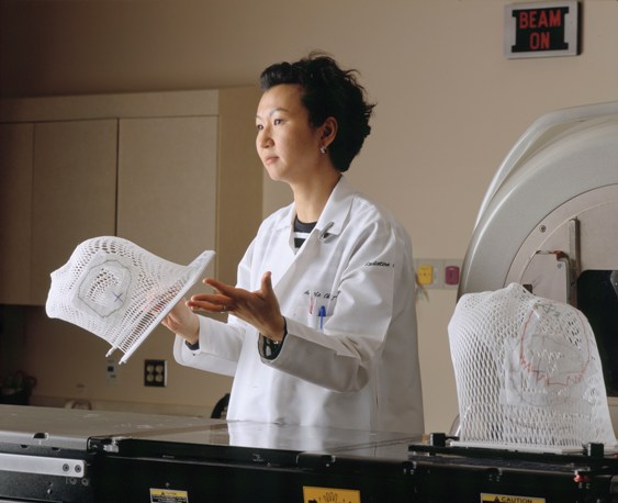
Attendant holding lattice face mask for positioning patient for radiation therapy
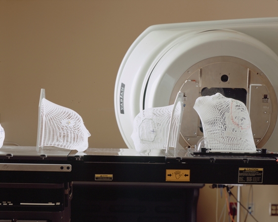
Lattice face masks sitting on patient bed prior to radiation therapy
 Print This Post