

Snapshot
Surveying Cells
Microglia scan the retina for damage and disease
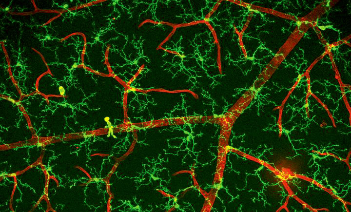
This image shows a sample of tissue from the light-sensitive retina in the back of the eye, as seen through a microscope. The green cells around the red blood vessels are called microglia. The blood vessels and microglia overlap in the yellow areas. Both the microglia and blood vessels are surrounded by retinal nerve cells that are not seen in this picture.
Microglia are an important part of the immune system in the retina. They survey the retina for signs of disease and cell damage by constantly moving their long, arm-like extensions back and forth in the space around blood vessels and nerve cells.
In eye diseases such as age-related macular degeneration and diabetic retinopathy, microglia pull in their extensions and change to a more rounded shape. They also move toward diseased areas of the retina.
Scientists are currently studying what happens when microglia migrate to and interact with diseased retina tissue. When they understand this process, researchers can potentially use microglia as targets for new eye disease treatments.
Image of a healthy mouse retina courtesy of Wai Wong, M.D., Ph.D., staff clinician and chief of the NEI Unit on Neuron-Glia Interactions in Retinal Disease.

