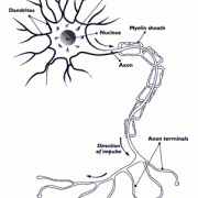
The brain is made up of billions of nerve cells, also known as neurons. Typically, a neuron contains three important parts (Figure 4): a central cell body that directs all activities of the neuron; dendrites, short fibers that receive messages from other neurons and relay them to the cell body; and an axon, a long single fiber that transmits messages from the cell body to the dendrites of other neurons or to body tissues, such as muscles. Although most neurons contain all of the three parts, there is a wide range of diversity in the shapes and sizes of neurons as well as their axons and dendrites.
Click the image to enlarge
Figure 4: Drawing of a neuron. The neuron shows the Dendrites, Cell Body, Nucleus, Myelin Sheath, Axon, Axon Terminals.
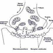
The transfer of a message from the axon of one nerve cell to the dendrites of another is known as neurotransmission. Although axons and dendrites are located extremely close to each other, the transmission of a message from an axon to a dendrite does not occur through direct contact. Instead, communication between nerve cells occurs mainly through the release of chemical substances into the space between the axon and dendrites (Figure 5). This space is known as the synapse. When neurons communicate, a message, traveling as an electrical impulse, moves down an axon and toward the synapse. There it triggers the release of molecules called neurotransmitters from the axon into the synapse. The neurotransmitters then diffuse across the synapse and bind to special molecules, called receptors, that are located within the cell membranes of the dendrites of the adjacent nerve cell. This, in turn, stimulates or inhibits an electrical response in the receiving neuron's dendrites. Thus, the neurotransmitters ac as chemical messengers, carrying information from one neuron to another.
Figure 5: Click the image to enlarge
There are many different types of neurotransmitters, each of which has a precise role to play in the functioning of the brain. Generally, each neurotransmitter can only bind to a very specific matching receptor. Therefore, when a neurotransmitter couples to a receptor, it is like fitting a key into a lock. This coupling then starts a whole cascade of events at both the surface of the dendrite of the receiving nerve cell and inside the cell. In this manner, the message carried by the neurotransmitter is received and processed by the receiving nerve cell. Once this has occurred, the neurotransmitter is inactivated in one of two ways. It is either broken down by an enzyme or reabsorbed back into the nerve cell that released it. The reabsorption (also known as re-uptake) is accomplished by what are known as transporter molecules (Figure 5). Transporter molecules reside in the cell membranes of the axons that release the neurotransmitters. They pick up specific neurotransmitters from the synapse and carry them back across the cell membrane and into the axon. The neurotransmitters are then available for reuse at a later time.
As noted above, messages that are received by dendrites are relayed to the cell body and then to the axon. The axons then transmit the messages, which are in the form of electrical impulses, to other neurons or body tissues. The axons of many neurons are covered in a fatty substance known as myelin. Myelin has several functions. One of its most important is to increase the rate at which nerve impulses travel along the axon. The rate of conduction of a nerve impulse along a heavily myelinated axon can be as fast as 120 meters/second. In contrast, a nerve impulse can travel no faster than about 2 meters/second along an axon without myelin. The thickness of the myelin covering on an axon is closely linked to the function of that axon. For example, axons that travel a long distance, such as those that extend from the spinal cord to the foot, generally contain a thick myelin covering to facilitate faster transmission of the nerve impulse. (Note: The axons that transmit messages from the brain or spinal co rd to muscles and other body tissues are what make up the nerves of the human body. Most of these axons contain a thick covering of myelin, which accounts for the whitish appearance of nerves.)


