
General Information About Adult Non-Hodgkin Lymphoma
Key Points for This Section
- Adult non-Hodgkin lymphoma is a disease in which malignant (cancer) cells form in the lymph system.
- There are many different types of lymphoma.
- Waldenström macroglobulinemia is a type of non-Hodgkin lymphoma.
- Age, gender, and a weakened immune system can affect the risk of adult non-Hodgkin lymphoma.
- Possible signs of adult non-Hodgkin lymphoma include fever, sweating, fatigue, and weight loss.
- Tests that examine the body and lymph system are used to help detect (find) and diagnose adult non-Hodgkin lymphoma.
- Certain factors affect prognosis (chance of recovery) and treatment options.
Adult non-Hodgkin lymphoma is a disease in which malignant (cancer) cells form in the lymph system.
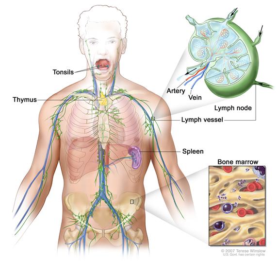
The lymph system is part of the immune system and is made up of the following:
- Lymph: Colorless, watery fluid that travels through the lymph system and carries white blood cells called lymphocytes. Lymphocytes protect the body against infections and the growth of tumors.
- Lymph vessels: A network of thin tubes that collect lymph from different parts of the body and return it to the bloodstream.
- Lymph nodes: Small, bean-shaped structures that filter lymph and store white blood cells that help fight infection and disease. Lymph nodes are located along the network of lymph vessels found throughout the body. Clusters of lymph nodes are found in the underarm, pelvis, neck, abdomen, and groin.
- Spleen: An organ that makes lymphocytes, filters the blood, stores blood cells, and destroys old blood cells. It is on the left side of the abdomen near the stomach.
- Thymus: An organ in which lymphocytes grow and multiply. The thymus is in the chest behind the breastbone.
- Tonsils: Two small masses of lymph tissue at the back of the throat. The tonsils make lymphocytes.
- Bone marrow: The soft, spongy tissue in the center of large bones. Bone marrow makes white blood cells, red blood cells, and platelets.
Because lymph tissue is found throughout the body, adult non-Hodgkin lymphoma can begin in almost any part of the body. Cancer can spread to the liver and many other organs and tissues.
Non-Hodgkin lymphoma in pregnant women is the same as the disease in nonpregnant women of childbearing age. However, treatment is different for pregnant women. This summary includes information on the treatment of non-Hodgkin lymphoma during pregnancy
Non-Hodgkin lymphoma can occur in both adults and children. Treatment for children, however, is different than treatment for adults. (See the PDQ summary on Childhood Non-Hodgkin Lymphoma Treatment for more information.)
There are many different types of lymphoma.
Lymphomas are divided into two general types: Hodgkin lymphoma and non-Hodgkin lymphoma. This summary is about the treatment of adult non-Hodgkin lymphoma. For information about other types of lymphoma, see the following PDQ summaries:
- Adult Acute Lymphoblastic Leukemia Treatment
- Adult Hodgkin Lymphoma Treatment
- AIDS-Related Lymphoma Treatment
- Chronic Lymphocytic Leukemia Treatment
- Hairy Cell Leukemia Treatment
- Mycosis Fungoides and the Sézary Syndrome Treatment
- Primary CNS Lymphoma Treatment
- Childhood Non-Hodgkin Lymphoma Treatment
Waldenström macroglobulinemia is a type of non-Hodgkin lymphoma.
Waldenström macroglobulinemia begins in a type of white blood cell called B lymphocytes. Certain B lymphocytes multiply out of control and make large amounts of a protein called monoclonal immunoglobulin M (IgM) antibody. High levels of IgM in the blood cause the blood to thicken and leads to many of the symptoms of Waldenström macroglobulinemia. Waldenström macroglobulinemia is also called lymphoplasmacytic lymphoma.
Age, gender, and a weakened immune system can affect the risk of adult non-Hodgkin lymphoma.
Anything that increases your risk of getting a disease is called a risk factor. Having a risk factor does not mean that you will get cancer; not having risk factors doesn’t mean that you will not get cancer. Talk with your doctor if you think you may be at risk. Risk factors for adult non-Hodgkin lymphoma include the following:
- Being older, male, or white.
- Having one of the following medical conditions:
- An inherited immune disorder (for example, hypogammaglobulinemia or Wiskott-Aldrich syndrome).
- An autoimmune disease (for example, rheumatoid arthritis, psoriasis, or Sjögren syndrome).
- HIV /AIDS.
- Human T-lymphotrophic virus type I or Epstein-Barr virus.
- A history of Helicobacter pylori infection.
- Taking immunosuppressant drugs after an organ transplant.
- Being exposed to certain pesticides.
- A diet high in meats and fat.
- Past treatment for Hodgkin lymphoma.
Possible signs of adult non-Hodgkin lymphoma include fever, sweating, fatigue, and weight loss.
These and other symptoms may be caused by adult non-Hodgkin lymphoma. Other conditions may cause the same symptoms. Check with your doctor if you have any of the following problems:
- Painless swelling in the lymph nodes in the neck, underarm, groin, or stomach.
- Fever for no known reason.
- Drenching night sweats.
- Feeling very tired.
- Weight loss for no known reason.
- Skin rash or itchy skin.
- Pain in the chest, abdomen, or bones for no known reason.
Symptoms of Waldenström macroglobulinemia depend on the part of the body affected. Most patients with Waldenström macroglobulinemia have no symptoms. Check with your doctor if you have any of the following problems:
- Feeling very tired.
- Headache.
- Easy bruising or bleeding, such a nosebleeds or bleeding from the gums.
- Vision changes, such as blurred vision or blind spots.
- Dizziness.
- Pain, tingling, or numbness, especially in the hands, feet, fingers, or toes.
- Confusion.
- Pain or a feeling of fullness below the ribs on the left side.
- Painless lumps in the neck, underarm, stomach, or groin.
- Weight loss for no known reason.
Tests that examine the body and lymph system are used to help detect (find) and diagnose adult non-Hodgkin lymphoma.
The following tests and procedures may be used:
- Physical exam and history: An exam of the body to check general signs of health, including checking for signs of disease, such as lumps or anything else that seems unusual. A history of the patient’s health habits and past illnesses and treatments will also be taken.
- Blood and urine immunoglobulin studies: A procedure in which a blood or urine sample is checked to measure the amounts of certain antibodies (immunoglobulins). In Waldenström macroglobulinemia, immunoglobulin M (IgM) and beta-2-microglobulin is measured. A higher- or lower-than-normal amount of these substances can be a sign of disease.
- Blood viscosity test: A procedure in which a blood sample is checked to see how “thick” the blood is. In Waldenström macroglobulinemia, when the amount of monoclonal immunoglobulin M (IgM) antibody in the blood becomes very high, the blood thickens and may cause symptoms.
- Flow cytometry: A laboratory test that measures the number of cells in a sample, the percentage of live cells in a sample, and certain characteristics of cells, such as size, shape, and the presence of tumor markers on the cell surface. The cells are stained with a light-sensitive dye, placed in a fluid, and passed in a stream before a laser or other type of light. The measurements are based on how the light-sensitive dye reacts to the light. This test is used to diagnose Waldenström macroglobulinemia.
- Bone marrow aspiration and biopsy: The removal of bone marrow, blood, and a small piece of bone by inserting a needle into the hipbone or breastbone. A pathologist views the bone marrow, blood, and bone under a microscope to look for signs of cancer.Enlarge
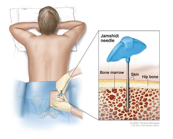 Bone marrow aspiration and biopsy. After a small area of skin is numbed, a Jamshidi needle (a long, hollow needle) is inserted into the patient’s hip bone. Samples of blood, bone, and bone marrow are removed for examination under a microscope.
 - Lumbar puncture: A procedure used to collect cerebrospinal fluid from the spinal column. This is done by placing a needle into the spinal column. This procedure is also called an LP or spinal tap. A pathologist views the cerebrospinal fluid under a microscope to look for signs of cancer.Enlarge
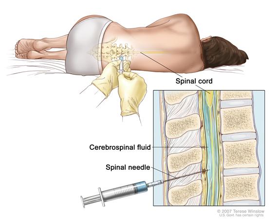 Lumbar puncture. A patient lies in a curled position on a table. After a small area on the lower back is numbed, a spinal needle (a long, thin needle) is inserted into the lower part of the spinal column to remove cerebrospinal fluid (CSF, shown in blue). The fluid may be sent to a laboratory for testing.
 - Lymph node biopsy: The removal of all or part of a lymph node. A pathologist views the tissue under a microscope to look for cancer cells. One of the following types of biopsies may be done:
- Excisional biopsy: The removal of an entire lymph node.
- Incisional biopsy: The removal of part of a lymph node.
- Core biopsy: The removal of part of a lymph node using a wide needle.
- Fine-needle aspiration (FNA) biopsy: The removal of tissue or fluid using a thin needle.
- Laparoscopy: A surgical procedure to look at the organs inside the abdomen to check for signs of disease. Small incisions (cuts) are made in the wall of the abdomen and a laparoscope (a thin, lighted tube) is inserted into one of the incisions. Other instruments may be inserted through the same or other incisions to perform procedures such as removing organs or taking tissue samples to be checked under a microscope for signs of disease.
- Laparotomy: A surgical procedure in which an incision (cut) is made in the wall of the abdomen to check the inside of the abdomen for signs of disease. The size of the incision depends on the reason the laparotomy is being done. Sometimes organs are removed or tissue samples are taken and checked under a microscope for signs of disease.
If cancer is found, the following tests may be done to study the cancer cells:
- Immunohistochemistry study: A laboratory test in which a substance such as an antibody, dye, or radioisotope is added to a sample of cancer tissue to test for certain antigens. This type of study is used to tell the difference between different types of cancer.
- Cytogenetic analysis: A laboratory test in which cells in a sample of tissue are viewed under a microscope to look for certain changes in the chromosomes.
- Immunophenotyping: A process used to identify cells, based on the types of antigens or markers on the surface of the cell. This process is used to diagnose specific types of leukemia and lymphoma by comparing the cancer cells to normal cells of the immune system.
Certain factors affect prognosis (chance of recovery) and treatment options.
The prognosis (chance of recovery) and treatment options depend on the following:
- The stage of the cancer.
- The type of non-Hodgkin lymphoma.
- The amount of lactate dehydrogenase (LDH) in the blood.
- The amount of beta-2-microglobulin in the blood (for Waldenström macroglobulinemia).
- The patient’s age and general health.
- Whether the lymphoma has just been diagnosed or has recurred (come back).
For non-Hodgkin lymphoma during pregnancy, the treatment options also depend on:
- The wishes of the patient
- Which trimester of pregnancy the patient is in.
Some types of non-Hodgkin lymphoma spread more quickly than others do. Most non-Hodgkin lymphomas that occur during pregnancy are aggressive. Delaying treatment of aggressive lymphoma until after the baby is born may lessen the mother's chance of survival. Immediate treatment is often recommended, even during pregnancy.
 Back to Top