
Print
 Download Reader
 Text
Text


Cell Images Aid Researchers, Educators and the Public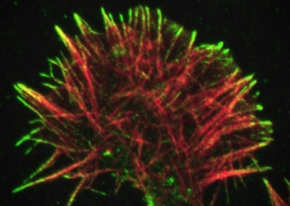 Image courtesy of Catherine Galbraith, James Galbraith. Published in Science, 2007, 315(5814):992-5. PMID: 17303755. Thanks to Recovery Act funding from the National Institutes of Health (NIH), the American Society for Cell Biology (ASCB) is making thousands of revealing, beautiful, and informative cell images and videos accessible worldwide to anyone with an Internet connection. From black-and-white historical cell images to video of an amoeba trying hard to ingest a mutant yeast cell, The Cell: An Image Library™ offers a carefully curated, searchable, annotated selection of images, videos, and animations of cell functions and interactions. While ASCB has long held the development of the library as a goal in its efforts to aid the scientific community and educators, its realization was impossible until the Society was awarded a Recovery Act grant for $2.5 million through the NIH. The grant enabled the Society to hire the necessary staff and consultants. For anyone interested in what the most basic unit of life looks like, the library provides a growing wealth of images, showing easily recognizable cell functions like egg fertilization. The library is designed to provide a valuable service to researchers and educators, and also to engage an interested public. Before the library’s creation, those looking for cell depictions were limited to images that had already been published in research journals, which are not organized for easy access. For example, if a researcher wanted to see a cell’s immune system process, she would need to go through back issues of journals to look for pictures that might have been previously published, reach out to colleagues to solicit their files, or go into the laboratory and create images of the cell process herself. Today, the scientist could bypass these time-consuming steps by simply querying the library. Additionally, if the scientist wanted to see a particular process in a specific organism or type of cell, she could quickly browse hundreds of other images to help illustrate her work. Many scientists are finding the library useful as a cataloguing system for their own collection of cell images. By making their work available to the library’s team of cell biologists for annotation and upload, researchers can eliminate sizable files in favor of a searchable database — all while showcasing their work and helping their colleagues, students, and others. The database also offers multiple depictions of the same subject. For a scientist who wants to learn about dendrite development, the library’s collection of more than 280 photos on the topic will provide an unparalleled education. Rather than trying to learn from an isolated image, seeing the same cell process depicted again and again helps users gain a more nuanced understanding of cell function. As the number and variety of images grow, the library holds great potential for being an educational resource for physicians. ASCB will eventually add images showing how cells respond to drug treatments, comparisons of healthy and damaged organ tissue, cancer cells, and other images that could aid in diagnosis or lead to other biomedical applications. In just six months, The Cell: An Image Library has had over 114,000 page views, with nearly 20,000 unique visitors clicking on the site. Visitors have come from 135 different countries, and ASCB anticipates that the site may be a great resource especially for scientists in the U.S. and abroad who lack funding and access to powerful microscopes. As the site grows toward its two-year goal of hosting 15,000 images, videos, and animations, ASCB continues its outreach to the scientific and educational communities to grow the collection and alert potential users that the resource is now available. |
Get Connected |